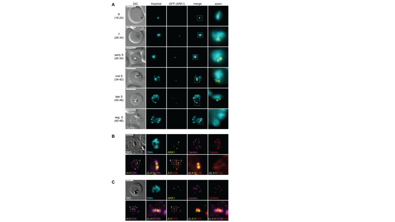
PfARK1 is expressed in a subset of nuclei during schizogony. (A) Localization of PfARK1–GFP throughout intra-erythrocytic asexual development as assessed by live cell fluorescence microscopy. Numbers in brackets indicate the age range (hours post invasion) of the synchronous parasite samples imaged at consecutive time points across intra-erythrocytic development. R, ring stage; T, trophozoite; S, schizont; seg., segmented. (B, C) Localization of PfARK1-GFP, centrin, and α/β-tubulin (B) or CENH3 (C) in schizonts as assessed by IFAs. Representative images are shown in each panel. DNA was stained with Hoechst (live) or 4′,6-diamidino-2-phenylindole (DAPI) (IFA). DIC, differential interference contrast. A1, PfARK1; CEN, centrin; TUB, α/β-tubulin; CH3, CENH3; Scale bar = 5 μWyss M, Thommen BT, Kofler J, Carrington E, Brancucci NMB, Voss TS. The three Plasmodium falciparum Aurora-related kinases display distinct temporal and spatial associations with mitotic structures in asexual blood stage parasites and gametocytes. mSphere. 2024 e0046524.
Other associated proteins
| PFID | Formal Annotation |
|---|---|
| PF3D7_0422300 | alpha tubulin 2, transcript expression profile microgametocyte |
| PF3D7_0903700 | alpha tubulin 1 |
| PF3D7_1008700 | tubulin beta chain |
| PF3D7_1333700 | histone H3-like centromeric protein CSE4 |
