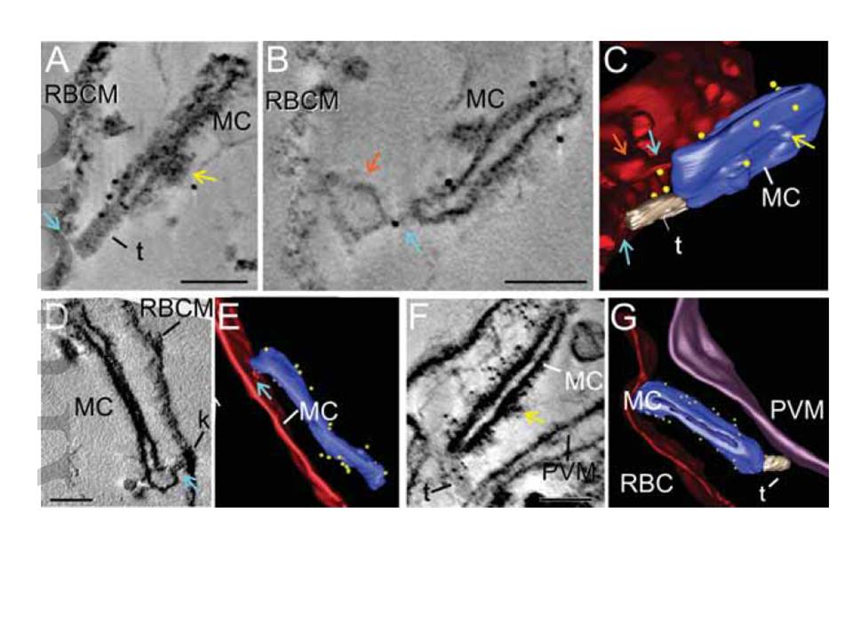
Immuno-electron tomography of Maurer's cleft-associated PfEMP1. EqtII-permeabilized PfEMP1B-GFP transfectants were labeled with antibodies recognizing GFP and prepared for electron tomography. Virtual sections (thickness: 24 nm A,B; 1.2 nm D; 1.5 nm F) from the tomograms and rendered models are presented. (A-C) A tether-like structure (t) connects a Maurer's cleft (MC) to the RBC membrane. (D, E) Region where the Maurer's cleft is closely opposed to the RBC membrane. (F,G) A tether-like structure (t) connects the Maurer's cleft (MC) to the PV membrane. The RBC membrane is rendered in red, the Maurer's clefts in blue, tethers in gray, the PV membrane in purple, and gold particles in yellow. A bulge in the RBC membrane is indicated with orange arrows. Regions that may represent RBC cytoskeleton extensions are indicated with aqua arrows. Thickening of the Maurer's cleft coat is observed in some regions and is indicated with yellow arrows. Scale bars = 100 nm.
McMillan PJ, Millet C, Batinovic S, Maiorca M, Hanssen E, Kenny S, Muhle RA, Melcher M, Fidock DA, Smith JD, Dixon MW, Tilley L. Spatial and temporal mapping of the PfEMP1 export pathway in Plasmodium falciparum. Cell Microbiol. 2013 15(8):1401-18PMID:
