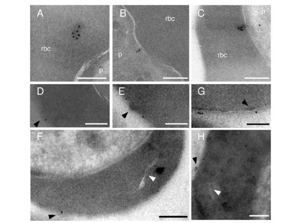
Ultrastructural analysis of miniPfEMP1 transport. The NTS-V5-TM-ATS-GFP or R29var-V5-GFP-TM-ATS parasite lines were analyzed by immunogold electron microscopy. A. NTS-V5-TM-ATS-GFP, immunogold particles were observed in association with an electron dense structure in the RBC cytoplasm. B. NTS-V5-TM-ATS-GFP localized close to the parasite plasma membrane. C. R29var-V5-GFP-TMATS immunogold particles were observed close to the PVM. D–H. R29varV5-GFP-TM-ATS parasites were labeled with anti-GFP, revealing miniPfEMP1 fusion proteins at the RBC surface at or near knob structures, as well as near a Maurer’s cleft in the RBC cytoplasm (H). Anti-GFP immunogold label is found at the red blood cell surface or in close proximity to Maurer’s clefts (H). Labeling with 10-nm immunogold was performed after cryosectioning (A to F and H) or prior to fixation with 2% glutaraldehyde (G). White arrowheads, Maurer’s clefts; black arrowheads, surface knobs of infected RBCs. Scale bars 200 nm, p parasite, rbc red blood cell cytoplasm.
Melcher M, Muhle RA, Henrich PP, Kraemer SM, Avril M, Vigan-Womas I, Mercereau-Puijalon O, Smith JD, Fidock DA. Identification of a Role for the PfEMP1 Semi-Conserved Head Structure in Protein Trafficking to the Surface of Plasmodium falciparum Infected Red Blood Cells. Cell Microbiol. 2010 12(10):1446-62 Copyright John Wiley & Sons Ltd. 2010.
