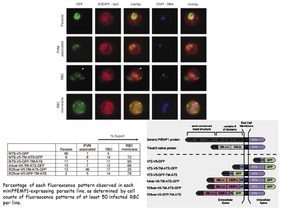
Localization of miniPfEMP1 proteins in the infected red blood cells by live cell fluorescence imaging. A. Characteristic fluorescence patterns observed in the miniPfEMP1-expressing parasite lines: 1st row: localization to the intracellular parasite; 2nd row: association with the parasite membrane/parasitophorous vacuole membrane (PVM); 3rd row: export into the RBC; 4th row: export to or near the RBC membrane. White arrowheads indicate GFP signal in lipid enclosed vesicles in the RBC cytoplasm. GFP fluorescence allowed us to identify several patterns of miniPfEMP1 trafficking: retention within the parasite (“Parasite”), retention at the PVM (“PVM associated”), export into the RBC cytoplasm (“RBC”), or localization at or near the RBC membrane (“RBC membrane”).
Melcher M, Muhle RA, Henrich PP, Kraemer SM, Avril M, Vigan-Womas I, Mercereau-Puijalon O, Smith JD, Fidock DA. Identification of a Role for the PfEMP1 Semi-Conserved Head Structure in Protein Trafficking to the Surface of Plasmodium falciparum Infected Red Blood Cells. Cell Microbiol. 2010 12(10):1446-62 Copyright John Wiley & Sons Ltd. 2010.
