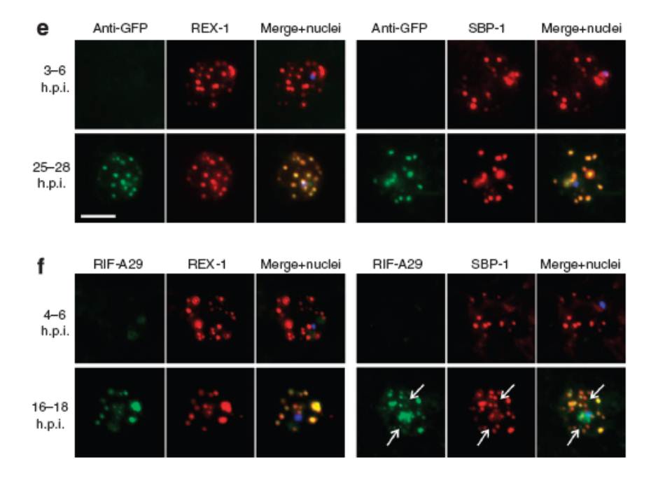
Export of membrane proteins is independent of cleft formation. IFA showing that in REX2-GFPcrt-expressing parasites (e) and 3D7 (f), early clefts contain REX1 and but not REX2-GFPcrt (e, upper panels) or RIFIN A29 (RIF-A29, f, upper panels). On progress of the culture to the trophozoite stage, the clefts containing REX1 and now also contain REX2-GFPcrt (e, lower panel) and RIFIN A29 (f, lower panel). Dots positive for RIFIN A29 only, found in ~50% of cells, are indicated by white arrows. Scale bars, 5 μm; h.p.i., hours post invasion. the clefts in early stages contained only the early markers (e,f). In contrast, later stages were positive for all four antigens (e,f). Crucially, in later stages all antigens occupied the same clefts (e,f), demonstrating that the clefts initially containing REX1 and SBP1 accumulated the other proteins later on. This establishes that export occurred without cleft formation.
Grüring C, Heiber A, Kruse F, Ungefehr J, Gilberger TW, Spielmann T. Development and host cell modifications of Plasmodium falciparum blood stages in four dimensions. Nat Commun. 2011 2:165.
Other associated proteins
| PFID | Formal Annotation |
|---|---|
| PF3D7_0501300 | skeleton-binding protein 1 |
| PF3D7_0935900 | ring-exported protein 1 |
| PF3D7_0936000 | ring-exported protein 2 |
