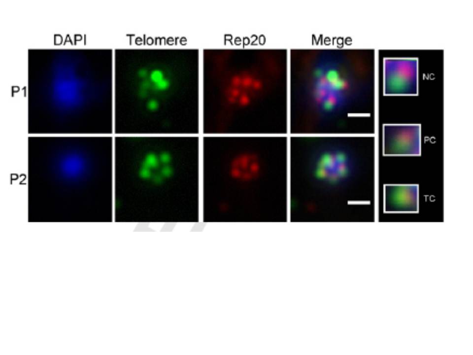
Examples of FISH images of P1- and P2-FISH of DAPI-stained nuclei (blue) hybridized with telomere (green) and Rep20 (red) Q1 probes and scored for colocalization (see Table 1). Bar, 1mm. Islets (far right) show examples of non-colocalization (NC), partial colocalization (PC) and total colocalization (TC) between both probes. P1-FISH increases the detection of telomeric clusters but causes their redistribution to central nuclear areas and reduces the colocalization index of telomeric and Rep20 probes. P1-FISH nuclei are almost twice as large and more variable in size than nuclei from P2-FISH.
Contreras-Dominguez M, Moraes CB, Dorval T, Genovesio A, Dossin Fde M, Freitas-Junior LH. A modified fluorescence in situ hybridization protocol for Plasmodium falciparum greatly improves nuclear architecture conservation. Mol Biochem Parasitol. 2010 173:48-52. Copyright Elsevier 2011
Other associated proteins
| PFID | Formal Annotation |
|---|---|
| Telomere | Telomere |
