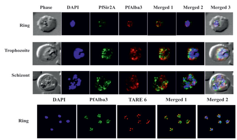
Upper panels: Plasmodium falciparum infected red blood cells were treated with DAPI (blue), mouse anti-PfSir2A antibody (secondary antibody Alexa Fluor 488, green) and rabbit anti-PfAlba3 antibody (secondary antibody Alexa Fluor 633, red), respectively. Overlay panels show the merged images in different combinations. PfAlba3 (red) and PfSir2A (green) fluorescence signals merge at several places, foremost at the peripheral region of the nucleus. However, co-localization was partial and not significant during schizont stage. Lower panel: FISH analysis combined with IF labeling of PfAlba3 to visualize telomeric region of chromosomes. Immunolocalization (IF) of PfAlba3 was performed at ring stage parasites using an Alexa 488-conjugated secondary antibody (green). FISH analysis was performed simultaneously. The nuclei were stained with DAPI (blue) and hybridized with TARE6 (Red) probe to visualize the chromosome ends on ring stage.
Goyal M, Alam A, Iqbal MS, Dey S, Bindu S, Pal C, Banerjee A, Chakrabarti S, Bandyopadhyay U. Identification and molecular characterization of an Alba-family protein from human malaria parasite Plasmodium falciparum. Nucleic Acids Res. 2011 40(3):1174-90.
Other associated proteins
| PFID | Formal Annotation |
|---|---|
| PF3D7_1006200 | DNA/RNA-binding protein Alba 3 |
| PF3D7_1328800 | transcriptional regulatory protein sir2a |
