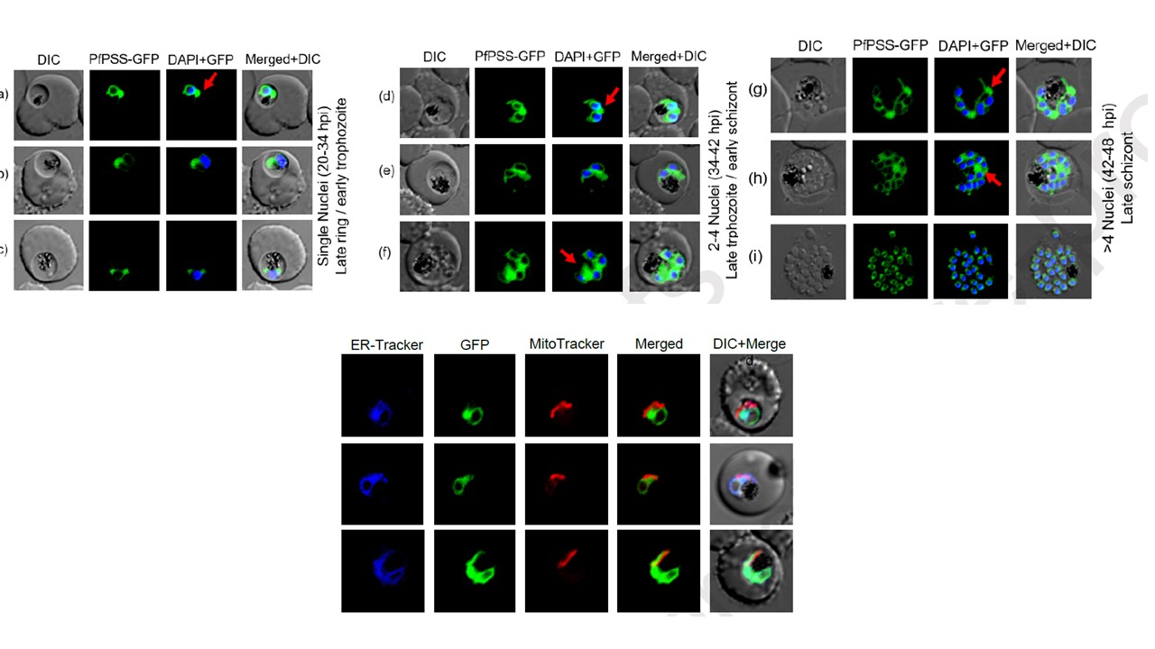
Dynamics of ER morphology in the intraerythrocytic stages of the P. falciparum parasite. (A) Live fluorescence microscopy images of PfPSS-GFP transgenic parasites at different developmental stages (hpi- hours post infection) of P. falciparum asexual life cycle. Parasite nuclei are stained with DAPI. Arrow denotes the peculiar morphological feature of ER, a bulging structure in cytoplasm observed in some parasite. Mitochondria is localized juxtaposed to the ER protrusions: (A) Live fluorescence microscopy images of PfPSS-GFP transgenic parasite stained with ER-Tracker Blue and MitoTracker Red. The PfPSS-GFP staining completely colocalizes with ER staining, whereas, ER protrusions in the cytosol are found to be juxtaposed to the mitochondrion forming a distinct ER mitochondria contact
Anwar O, Islam M, Thakur V, Kaur I, Mohmmed A. Defining ER-mitochondria contact dynamics in Plasmodium falciparum by targeting component of phospholipid synthesis pathway, Phosphatidylserine synthase (PfPSS). Mitochondrion. 2022 :S1567-7249(22)00046-0. PMID: 35623558.