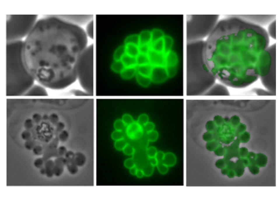
Confocal fluorescence microscopy images of schizont stage transfected 3D7 P. falciparum-infected RBCs expressing a GFP chimera directed to the parasite plasma membrane. DIC image, the GFP fluorescence signal and an overlay of a P. falciparum 92 cysteine-rich surface protein-GFP transfectant. Scale bar = 5 µm. Pf92 localizes to the merozoite surface and is a putative GPI-anchored protein distributed evenly over the parasite surface.
Reprinted from with permission from Elsevier: Tilley L, McFadden G, Cowman A, Klonis N. Illuminating Plasmodium falciparum-infected red blood cells. Trends Parasitol. 2007 23:268-77.
PubMed Article: Illuminating Plasmodium falciparum-infected red blood cells