EB1 like ARK2 associates with spindle and kinetochore during male
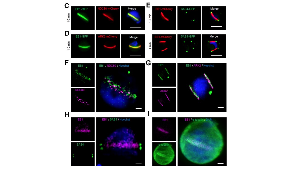
gametogony. (A) Live cell imaging of EB1-GFP (green) showing its location on spindles and spindle poles. DNA is stained with Hoechst dye (blue); scale bar = 5. μm. (B) ChIP-seq analysis of EB1-GFP profiles for all 14 chromosomes showing its centromeric binding. Signals are plotted on a normalized read per million (RPM) basis. Red lines at the top indicate the ends of chromosomes; circles on the bottom indicate centromere locations. NDC80-GFP was used as a positive control and IgG was used as a negative control. (C) Live cell imaging showing the location of EB1-GFP (green) and kinetochore marker NDC80-mCherry (red) in a gametocyte activated for 1-2 min. DNA is stained with Hoechst dye (blue); scale bar = 5 µm. (D) Live cell imaging showing the location of EB1-GFP (green) and ARK2-mCherry (red) in a gametocyte activated for 1-2 min. DNA is stained with Hoechst dye (blue); scale bar = 5 µm. (E) Live cell imaging showing the location of EB1-mCherry (red) and basal body marker SAS4-GFP (green) in gametocytes activated for 1 to 2 min (upper panel) and 4 min (lower panel). DNA is stained with Hoechst dye (blue); scale bar = 5 µm. (F) 3D-SIM image showing location of EB1 (green) and NDC80 (purple) in gametocyte activated for 1 min. DNA is stained with Hoechst dye (blue); scale bar = 1 μm. (G) 3D-SIM image showing location of EB1 (green) and ARK2 (purple) in gametocyte activated for 3 to 4 min. DNA is stained with Hoechst dye (blue); scale bar = 1 μm. (H) 3D-SIM image showing location of EB1 (purple) and cytoplasmic SAS4 (green) in gametocyte activated for 1 min. DNA is stained with Hoechst dye (blue); scale bar = 1 μm. (I) STED confocal microscopy showing co-localization of EB1 (purple) and α-tubulin (green) at spindle but not with cytoplasmic microtubules in gametocytes activated for 1 min. DNA is stained with SiR DNA (blue); scale bar = 1 μm.
Zeeshan M, Rea E, Abel S, Vukušić K, Markus R, Brady D, Eze A, Raspa R, Balestra A, Bottrill AR, Brochet M, Guttery DS, Tolić IM, Holder AA, Roch KGL, Tromer EC, Tewari R. Plasmodium ARK2-EB1 axis drives the unconventional spindle dynamics, scaffold formation and chromosome segregation of sexual transmission stages. bioRxiv [Preprint]. 2023 31:2023.01.29.526106
Other associated proteins
| PFID | Formal Annotation |
|---|---|
| PF3D7_0309200 | serine/threonine protein kinase ark2, putative |
| PF3D7_0616200 | kinetochore protein NDC80, putative |
| PF3D7_1458500 | spindle assembly abnormal protein 4, putative |