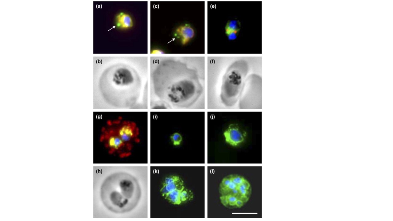
PfPDI localization in different parasite erythrocytic stages and co-localization with markers of subcellular compartments. (a,b) Immunostaining with antisera directed against rPfPDI (green) and PfBIP (red) and the corresponding phase contrast image of a trophozoite. (c,d) Immunostaining with antisera directed against rPfPDI (green) and PfERC (red) and the corresponding phase contrast image of a trophozoite. (e,f) Immunostaining with antisera directed against rPfPDI (green) and PfERD2 (red) and the corresponding phase contrast image of a young schizont. (g,h) Immunostaining with antisera directed against rPfPDI (green) and Pf332 (red) and the corresponding phase contrast image of an erythrocyte infected by two young trophozoites. Regions of total overlapping of both proteins appear a yellow-orange color, DNA staining by DAPI appears in blue color. Arrows indicate the absence of co-localization. (i–l) Overlaid images of parasites immunostained with antiserum directed against rPfPDI (green) and incubated with the nuclear dye DAPI (blue). Parasite stages were: ring (i), old trophozoites (j), schizont (k) and mature schizont with individualized merozoites (l). Bar: 5 μm.
Mouray E, Moutiez M, Girault S, Sergheraert C, Florent I, Grellier P. Biochemical properties and cellular localization of Plasmodium falciparum protein disulfide isomerase. Biochimie. 2007 89(3):337-46.
Other associated proteins
| PFID | Formal Annotation |
|---|---|
| PF3D7_0827900 | protein disulfide isomerase |
| PF3D7_1108600 | endoplasmic reticulum-resident calcium binding protein |
| PF3D7_1149000 | antigen 332, DBL-like protein |
| PF3D7_1353600 | ER lumen protein retaining receptor |