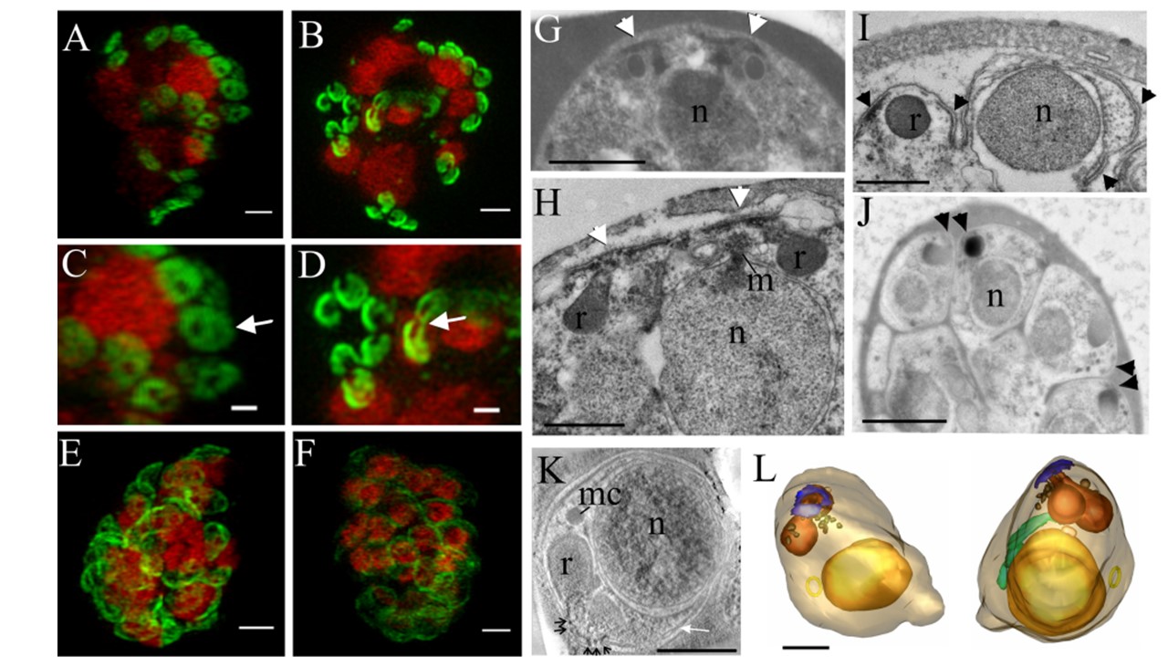
The IMC forms around the apical pore of developing merozoites. 3D-SIM was performed on parasites expressing PfGAP50-GFP (green) labeled with DAPI nuclear stain (depicted in red). (A and C) Schizont with eight nuclei, each with two associated PfGAP50-GFPcontaining llipsoids. Each ellipsoid has two unlabeled “pores” (arrow). (B and D) A schizont with eight nuclei that are undergoing division showing the PfGAP50-GFP-containing structures separating into claw-like structures (arrow). (E and F) Mature schizonts in which the PfGAP50-GFP fluorescence is more evenly distributed around the periphery of the parasite. (G and H) Transmission EM (TEM) images of early schizonts with electron-dense apical prominences with overlying membrane caps (arrowheads) forming in pairs on either side of a nucleus (n). The apical caps are connected to the underlying electron-dense rhoptries (r) and form close to a mitotic spindle (m). (I) TEM image of a maturing schizont showing invagination of the parasite plasma membrane around the daughter cells and separation of the apical caps (arrowheads). (J) TEM image of a mature schizont showing electron-dense structures at the apical ends of closely apposed daughter cells (arrowheads). (K) Selected virtual section (22 nm) through a tomogram showing a merozoite developing within a schizont. The electron-dense polar rings are indicated with black arrows, and the IMC with a white arrow. A microneme (mc) is visible. (L) Rendered tomographic reconstructions of merozoites, generated from serial sections through the schizont shown in panel K, showing the nucleus in yellow, rhoptries in burnt orange, dense granules in brown, the polar rings in blue, the mitochondrion in green, and a cytostomal ring as a yellow circle. Bars 1 mm (A to F, G, and J) and 500 nm (H, I, K, and L)