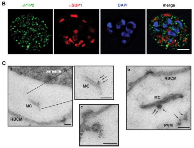
Localization of PfPTP2 in CS2idhfrPTP2/HA-infected RBCs. (B) First panel, PfPTP2 (green); second panel, SBP1 (Maurer’s cleft marker; red); third panel, DAPI (blue); fourth panel, all panels merged. (C) Immuno-EM of CS2idhfrPTP2/HA-infected RBCs after treatment with equinotoxin II. MC, Maurer’s cleft; RBCM, red blood cell plasma membrane; PVM, parasitophorous vacuole membrane. a, the side panel shows higher magnification of Maurer’s cleft; arrows point to budding vesicle where PfPTP2 is localized. b, arrows point to PfPTP2 on budding vesicle and membrane material. c, an example of budding vesicle with PfPTP2 localization. PfPTP2-labeled material in host cell cytoplasm was derived from Maurer’s clefts .
Regev-Rudzki N, Wilson DW, Carvalho TG, Sisquella X, Coleman BM, Rug M, Bursac D, Angrisano F, Gee M, Hill AF, Baum J, Cowman AF. Cell-Cell Communication between Malaria-Infected Red Blood Cells via Exosome-like Vesicles. Cell. 2013 May 23;153(5):1120-33.
Other associated proteins
| PFID | Formal Annotation |
|---|---|
| PF3D7_0731100 | gametocyte exported protein 11 EMP1-trafficking protein |