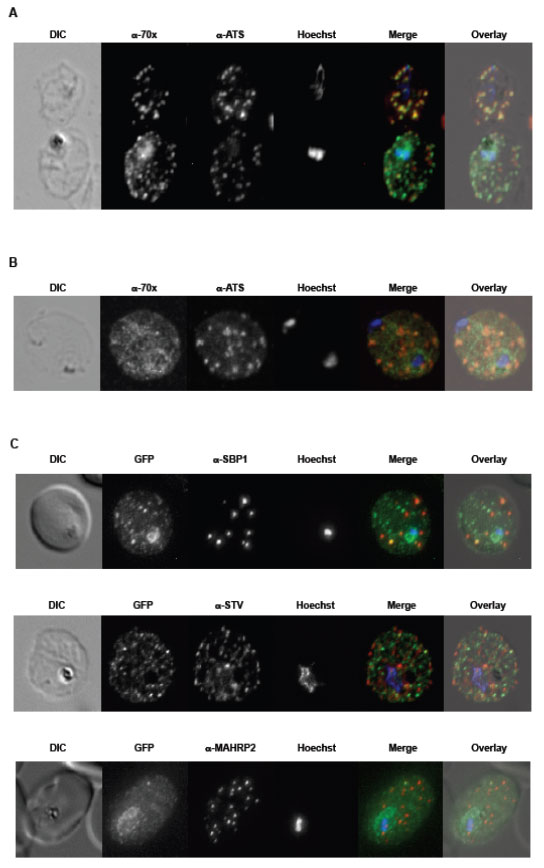
A. tage dependent co-localization of PfHsp70-x and PfEMP1. Upper infected cell (ring stage) shows a higher degree of signal colocalization than lower infected cell (trophozoite). B. ndirect immuno-fluorescence of PfHsp70-x and PfEMP1 using confocal microscopy. In merge and overlay: Green, α-70x; Red, α-ATS; Blue, Hoechst. C. ndirect immuno-fluorescence localization of PfHsp70-x using anti-SBP1, anti-MAHRP2 and anti-STEVOR (STV) antisera in cells infected with 3D7GFP:70x. In merge and overlay images: Green, GFP; red, specific antisera; Blue, Hoechst. No co-localization can be observed between the GFP and antibody-specific signals.
Külzer S, Charnaud S, Dagan T, Riedel J, Mandal P, Pesce ER, Blatch GL, Crabb BS, Gilson PR, Przyborski JM. Plasmodium falciparum-encoded exported hsp70/hsp40 chaperone/co-chaperone complexes within the host erythrocyte. Cell Microbiol. 2012 14(11):1784-95
Other associated proteins
| PFID | Formal Annotation |
|---|---|
| PF3D7_0501300 | skeleton-binding protein 1 |
| PF3D7_1040200 | PIR protein stevor |
| PF3D7_1353200 | membrane associated histidine-rich protein 2 |