1. BFA-treated (a) and control parasites in presence of ethanol (c) were analysed by immunofluorescence by using anti-MAP1 antibodies to detect PfA-M1 (red) and a mouse anti-exp2 serum as positive control (green). Nuclei, stained by using Hoechst 33342 appear in blue Corresponding phase contrasts are in (b, d). Scale bar, 5μm. In the control exp-2 was targeted as expected to the PVM and PfA-M1 was diffuse in the parasite (but outside the FV)
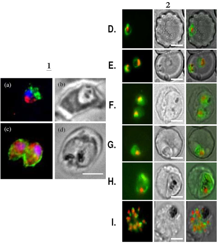
2. Live imaging of FcB1 parasites transfected with a construct allowing expression of PfA-M1-GFP chimera confirm its marginal association with the food vacuole. Live imaging of transfected parasites showing: D, E. ring stages. F, G, trophozoite stages; H. young schizont. I. segmenting schizont. First column: GFP (green) and nuclei (Hoechst 33342); second column: phase contrast; third column: overlay. Scale bar, 5μm.
Azimzadeh O, Sow C, Geze M, Nyalwidhe J, Florent I. Plasmodium falciparum PfA-M1 aminopeptidase is trafficked via the parasitophorous vacuole and marginally delivered to the food vacuole. Malar J. 2010 9:189:9