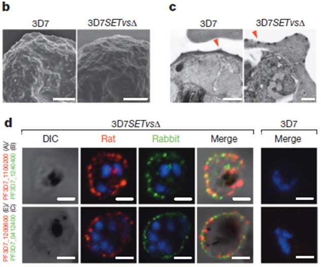
b and c: Electron microscopy of gelatin-selected 3D7 and 3D7SETvsD iRBCs. Typical knobs in scanning electron microscopy (b) and transmission electron microscopy (c) pictures are indicted by red arrowheads. d, Live-cell IFA using rat and rabbit antisera to various PfEMP1 proteins to detect co-expression of different PfEMP1 proteins on the surface of 3D7SETvsD (P. falciparum 3D7 lacking the SET2 gene) iRBCs. Wild-type 3D7 iRBC is shown to the right. No staining is seen. DAPI is used to mark the parasite nucleus. Types of var genes are shown in parentheses. Scale bars, 1 mm (b), 0.5 mm (c) and 1.5 mm (d). Knock out of PfSET2 led to the expression of virtually all var genes in the ring stage. By contrast, knockout of any other PfSET or PfHKDM genes did not alter the transcription of the var gene family in 3D7.
Jiang L, Mu J, Zhang Q, Ni T, Srinivasan P, Rayavara K, Yang W, Turner L, Lavstsen T, Theander TG, Peng W, Wei G, Jing Q, Wakabayashi Y, Bansal A, Luo Y, Ribeiro JM, Scherf A, Aravind L, Zhu J, Zhao K, Miller LH. PfSETvs methylation of histone H3K36 represses virulence genes in Plasmodium falciparum. Nature. 2013 499(7457):223-7.