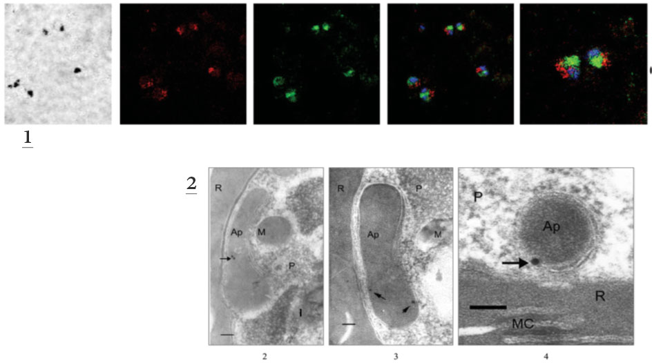
1. Analysis of the co-localization of PfFC with PfHsp60 using FITC-conjugated (green) secondary antibodies for the former and TRITC-conjugated (red) secondary antibodies for the latter; 1, bright field (dark spots represent the hemozoin pigment); 2, red fluorescence in the parasite due to PfHsp60; 3, green fluorescence in the parasite due to PfFC; 4, merge of 2 and 3 along with Hoescht stain (blue) showing lack of co-localization; 5, projection of two parasites showing lack of co-localization.
2. Three different fields showing discrete gold particles in typical apicoplast structures with multiple membranes. 2 part of the whole parasite; 3, elongating apicoplasts in a later-stage trophozoite; 4, a circular apicoplast in an early-stage tropozoite. The signals are localized close to the membrane. Scale bar, 100 μm. AP, apicoplast; M, mitochondrion; R, red cell; MC, Maurer’s cleft; P, parasite; Pv, parasite food vacuole; Pc, parasite cytosol.
Reproduced with permission, from Varadharajan S, Sagar BK, Rangarajan PN, Padmanaban G. Localization of ferrochelatase in Plasmodium falciparum. Biochem J. 2004 384:429-36. © the Biochemical Society