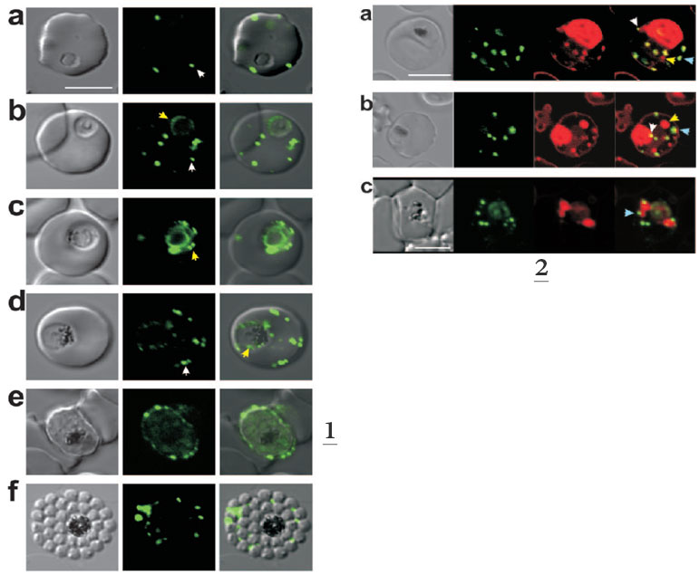
1. Expression of MAHRP11-249-GFP at different stages of the intraerythrocytic cycle of P. falciparum. The images represent a DIC image, the GFP fluorescence signal, and an overlay of these images. Ring and trophozoite stage parasites (a to d) show puncta of fluorescence in the RBC cytoplasm which appear to represent peripheral Maurer’s clefts (white arrows). Some cells (c) show a ring of “beads” of fluorescence around the PV. These foci may represent nascent Maurer’s clefts (yellow arrows). Mature schizont-stage parasites (e) show flattening of the peripheral Maurer’s clefts against the RBC membrane.
2. Dual labeling of MAHRP11-249-GFP ransfectants with BODIPY-ceramide. The images represent (from left to right) a DIC image, GFP fluorescence, BODIPY-ceramide fluorescence, and an overlay of the GFP (green) and BODIPY-ceramide (red) images. The parasite membranes are intensely labeled with the lipid probe. Some extensions of the PV membrane are dotted with foci of MAHRP1-GFP (white arrows). Some of the BODIPY-labeled structures (probably TVN extensions and buds) are not labeled with GFP (yellow arrows), while others (presumably Maurer’s clefts) are labeled with GFP (blue arrows). Bar, 5 mm.
Spycher C, Rug M, Klonis N, Ferguson DJ, Cowman AF, Beck HP, Tilley L. Genesis of and trafficking to the Maurer's clefts of Plasmodium falciparum-infected erythrocytes. Mol Cell Biol. 2006 26:4074-85.