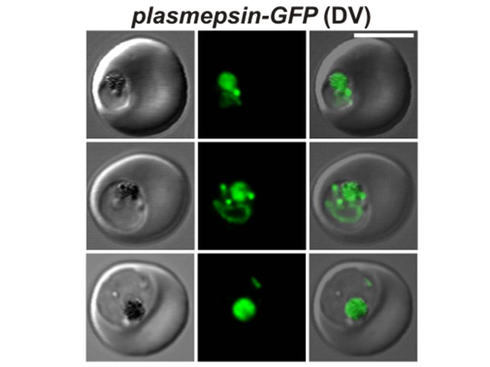
Confocal fluorescence microscopy images of transfected 3D7 P. falciparum-infected RBCs expressing a GFP chimera directed to the digestive vacuole. DIC image, the GFP fluorescence signal, and an overlay of a P. falciparum plasmepsin II-GFP transfectant. Scale bar = 5 µm. En route to the digestive vacuole during the ring and trophozoite stages plasmepsin-GFP is observed in the endoplasmic reticulum and in cytosome-derived vesicles (top and middle rows). In the late stages of parasite development, the chimera accumulates in the digestive vacuole as shown in the bottom row.
Tilley L, McFadden G, Cowman A, Klonis N. Illuminating Plasmodium falciparum-infected red blood cells. Trends Parasitol. 2007 23:268-77. PMID: 17434344
PubMed Article: Illuminating Plasmodium falciparum-infected red blood cells