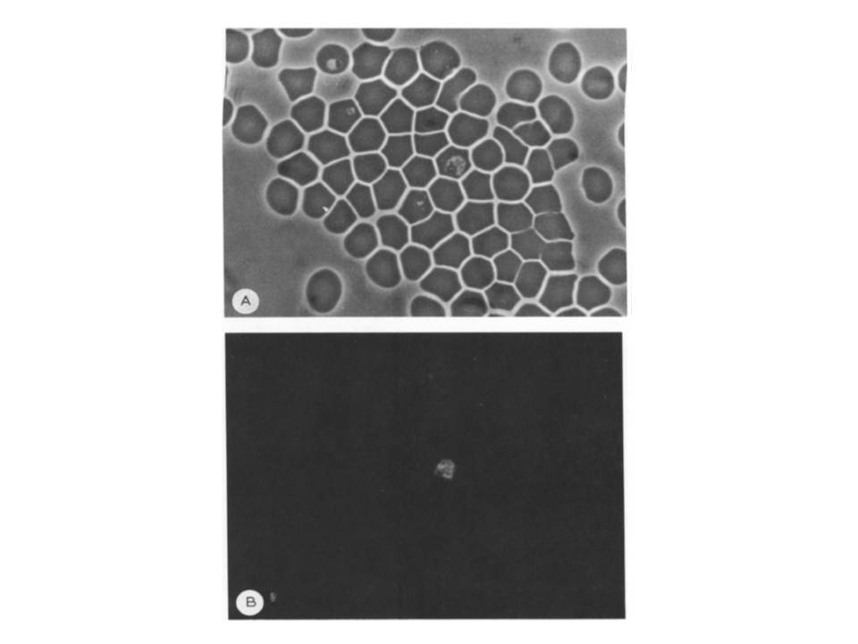
Indirect immunofluorescence of W2 infected erythrocytes with anti-pfmdr2 fusion protein. The slides containing fixed parasites were prepared from asynchronized W2 cultures as described in Materials and Methods. The cells were fixed with acetone and reacted with a rabbit anti-pfmdr2 fusion protein (dilution 1/50) followed by FITC-conjugated goat anti-rabbit IgG. (A) Light Microscopy. The arrow indicates a ring stage parasite. The trophozoite forms of infected erythrocytes can be identified by the hemozoin granules (dark crystals) within the digestive vacuole. (B) Fluorescent micrograph. The anti-pfmdr2 antibody only reacted with the trophozoite stage. the antibody staining appears to have a vacuolar localization.
Zalis MG, Wilson CM, Zhang Y, Wirth DF. Characterization of the pfmdr2 gene for Plasmodium falciparum. Mol Biochem Parasitol. 1993 62:83-92.