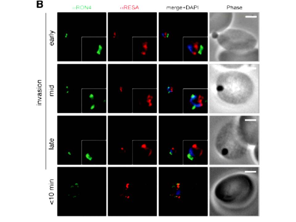
Wide field IFA time course of invasion using anti-RESA/PfRON4. Scale bar = 2.0 μm. Pre-egress shows standard wide-field image. Invasion images show IFA with deconvolution (single slice). RESA is retained within the merozoite body, often at the apical end but frequently basal to rhoptries and micronemes (RESA early). RESA, in contrast, retained its position inside the merozoite, establishing that release of dense granule proteins likely occurs postinvasion (RESA mid). RESA, localization became markedly more peripheral and evenly distributed around the merozoite immediately after invasion (RESA late), supporting the notion that dense granules are released after invasion at the merozoite plasma membrane.
Riglar DT, Richard D, Wilson DW, Boyle MJ, Dekiwadia C, Turnbull L, Angrisano F, Marapana DS, Rogers KL, Whitchurch CB, Beeson JG, Cowman AF, Ralph SA, Baum J. Super-resolution dissection of coordinated events during malaria parasite invasion of the human erythrocyte. Cell Host Microbe. 2011 9:9-20.
Other associated proteins
| PFID | Formal Annotation |
|---|---|
| PF3D7_1116000 | rhoptry neck protein 4 |