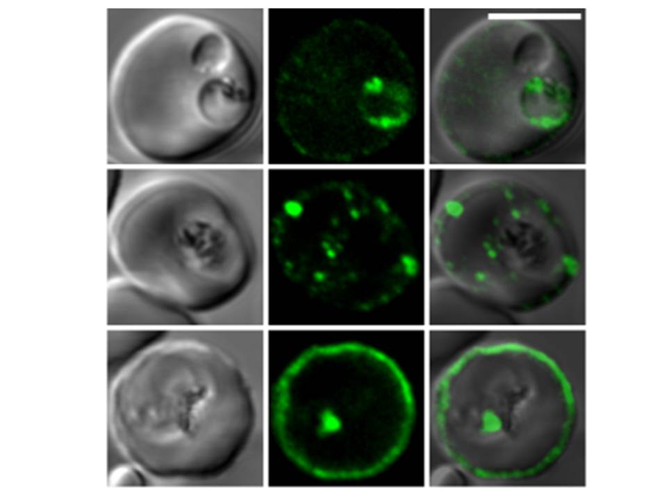
Confocal fluorescence microscopy images of transfected 3D7 P. falciparum-infected RBCs expressing a GFP chimera directed to the red blood cell membrane skeleton. DIC image, the GFP fluorescence signal and an overlay of a P. falciparum erythrocyte membrane protein-31-500 -GFP transfectant. Scale bar = 5 µm.
PfEMP3 is present in the parasite's endomembrane system in the ring stage (top row), and with the Maurer’s clefts (middle row) and red blood cell membrane skeleton (bottom row) in more mature stage parasites.
Tilley L, McFadden G, Cowman A, Klonis N. Illuminating Plasmodium falciparum-infected red blood cells. Trends Parasitol. 2007 23:268-77. Copyright Elsevier 2009.
PubMed Article: Illuminating Plasmodium falciparum-infected red blood cells