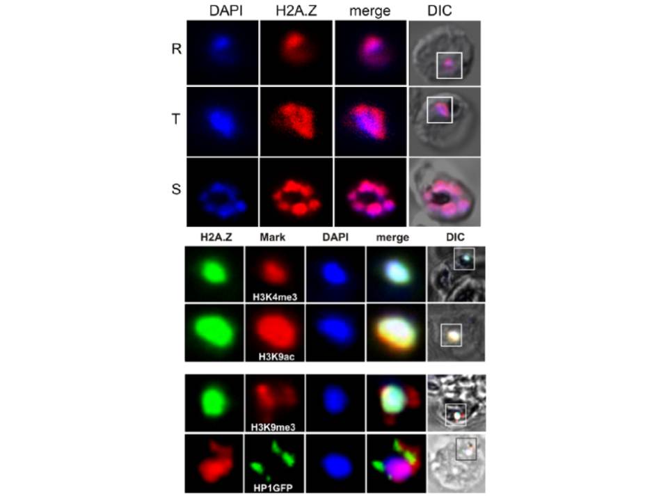
Upper panel: PfH2A.Z is expressed in the nucleus throughout asexual differentiation. Full length PfH2A.Z was expressed as a GSTfusion protein in E. coli and used to immunize rabbits. Nuclear localization of PfH2A.Z is shown by indirect immunofluorescence analysis and confocal microscopy of fixed 3D7 parasites using anti-PfH2A.Z antibodies. DNA was visualized with DAPI. R = ring stage, T =trophozoite stage, S = schizont stage. DIC = differential interference contrast. An area stained with the DNA dye DAPI but devoid of PfH2A.Z labelling was consistently observed, indicating that PfH2A.Z is enriched towards one side of the nucleus.
Lower panel: Colocalization analysis shows PfH2A.Z overlap with the euchromatin marks H3K9ac and H3K4me3 but not with the subtelomeric heterochromatin marks H3K9me3 and HP1. PfH2A.Z, H3K4me3, H3K9ac and H3K9me3 were detected with specific antibodies.
Petter M, Lee CC, Byrne TJ, Boysen KE, Volz J, Ralph SA, Cowman AF, Brown GV, Duffy MF. Expression of P. falciparum var Genes Involves Exchange of the Histone Variant H2A.Z at the Promoter. PLoS Pathog. 2011 7(2):e1001292. 17.