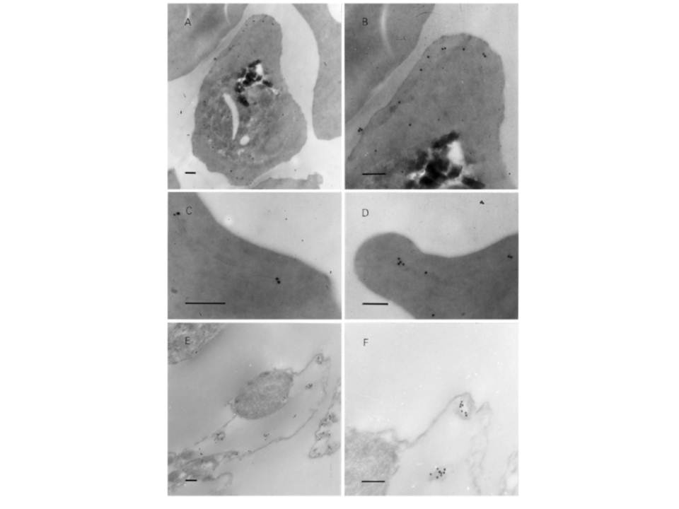
Post-embedding immunoelectron microscopy using 6B4 monoclonal. Panoramic view of an infected erythrocyte (A) and higher magnification images (B–D) showing the preferential location of gold particles over Maurer’s clefts. Panels (E) and (F) arephotographs of ‘parasite ghosts’ probed with 6B4, showing the reaction of the monoclonal antibody over cytoplasmic vesicles. Bar indicates 0.2 mm. Immunoelectron microscopy demonstrated that the monoclonal antibody reacts with cytoplasmic vesicles of Plasmodium falciparum infected erythrocyte referred to as Maurer’s clefts. 6B4 recognized 50 and 41 kDa antigens. The 50 kDa antigen is most probably PfSBP1.
Martinez SL, Clavijo CA, Winograd E. Identification of peripheral membrane proteins associated with the tubo-vesicular network of Plasmodium falciparum infected erythrocytes. Mol Biochem Parasitol. 1998 91:273-80. Copyright Elsevier 2009.