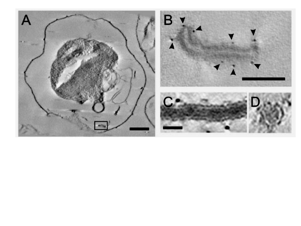
Electron tomography of individual Maurer’s cleft bodies, tubules and vesicles in a Plasmodium falciparum strain 3D7-infected red blood cell (RBC). (A) A virtual section with an individual Maurer’s cleft marked with a rectangle, which is depicted at higher resolution in (B). The presence of the gold particles (arrowheads) indicates labelling with antibodies recognising the Maurer’s cleft resident, skeleton-binding protein-1 (SBP1). (C and D) Virtual sections showing transverse (C) and cross-section (D) views through a tubular structure. Scale bars: (A) 1 mm; (B) 100 nm; (C and D) 25 nm..
Hanssen E, Carlton P, Deed S, Klonis N, Sedat J, Derisi J, Tilley L. Whole cell imaging reveals novel modular features of the exomembrane system of the malaria parasite, Plasmodium falciparum. Int J Parasitol. 2010 40:123-134. Copyright Elsevier 2010.