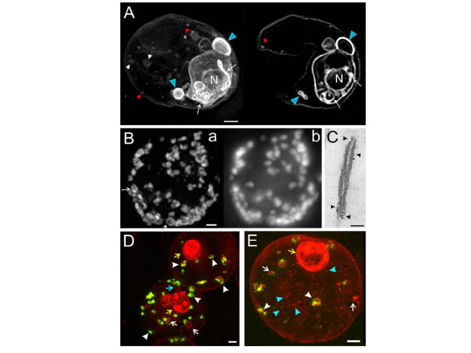
Three-dimensional structured illumination microscopy and immuno-electron microscopy (EM) of Plasmodium falciparum-infected red blood cells (RBCs). (A) 3D7 -infected RBCs (trophozoite stage) were labelled with the membrane probe, BODIPY-ceramide, and examined without fixation. Three Z-stacks (0.125 mm section spacing) were recorded. A projection image and a representative virtual section are presented. Features of the parasite’s endomembrane system (white arrows) and parasitophorous vacuole (PV) membrane/ tubulovesicular network (TVN) (blue arrowheads) are strongly labelled while the membranous features in the RBC cytoplasm (white/red arrowheads) are less strongly labelled. (B and C) RBCs infected with trophozoite stage 3D7 Ring Exported Protein-1-GFP (3D7_REX1-GFP) transfectants were permeabilized with Equinatoxin II, and labelled with anti-GFP antiserum followed by (B) AlexaRed-anti-IgG or (C) 6 nm gold protein A. (B) Z-stacks presented as the ‘‘raw” projection (b) or used to compute a structured illumination microscopy (SIM) image (a). A restricted pattern is revealed in some of the Maurer’s clefts (arrows). (C) Samples were prepared for EM. (D and E) RBCs infected with early trophozoite stage 3D7_REX1-GFP transfectants were labelled with the membrane probe, BODIPY-ceramide, and fixed with paraformaldehyde. REX1-GFP is concentrated in punctuate (green fluorescence) in the RBC cytoplasm; these structures represent Maurer’s clefts. The BODIPY-ceramide (red fluorescence) is concentrated in the parasite (yellow arrows) and in extensions of the parasite into the RBC cytoplasm (blue arrow) as well as in separate structures in the RBC cytoplasm. Some of the membrane features in the RBC cytoplasm contain REX1-GFP (white arrowheads) while others do not (white arrows). Small vesicle-like structures are observed (E, blue arrowheads). Scale bars: (A, B, D, and E) 1 mm; (C) 50 nm.
Hanssen E, Carlton P, Deed S, Klonis N, Sedat J, Derisi J, Tilley L. Whole cell imaging reveals novel modular features of the exomembrane system of the malaria parasite, Plasmodium falciparum. Int J Parasitol. 2010 40:123-34.Copyright Elsevier 2010.