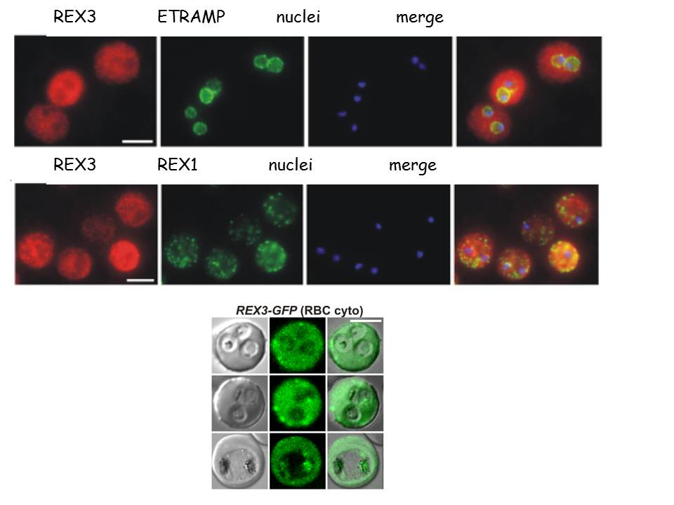
Upper row: Location of REX3 in IRBCs fixed with 4% formaldehyde/0.005% glutaraldehyde. IFAs with 3D7 parasites expressing a myc-tagged ETRAMP4 showed that REX3 (red; Texas Red) occurs as a uniform stain outside of the PVM (ETRAMP4myc in the PVM detected by a rabbit anti-myc antibody; green, Cy2).
Second row: REX3 (red; Texas Red) did not colocalize with the Maurer’s cleft protein REX1 (green; Cy2) in 3D7 parasites. Bars, 5 mm.
Lower panel: Confocal fluorescence microscopy images of transfected 3D7 P. falciparum-infected RBCs. REX3 is expressed in its early ring stages (top row) and is directed to the bulk compartment of the host cell cytoplasm. Trophozoites and schizonts (middle and bottom rows) display a uniform fluorescence pattern with some puncta.
Tilley L, McFadden G, Cowman A, Klonis N. Illuminating Plasmodium falciparum-infected red blood cells. Trends Parasitol. 2007 23:268-77.
Other associated proteins
| PFID | Formal Annotation |
|---|---|
| PF3D7_0423700 | early transcribed membrane protein 4 |
| PF3D7_0936300 | ring-exported protein 3 |