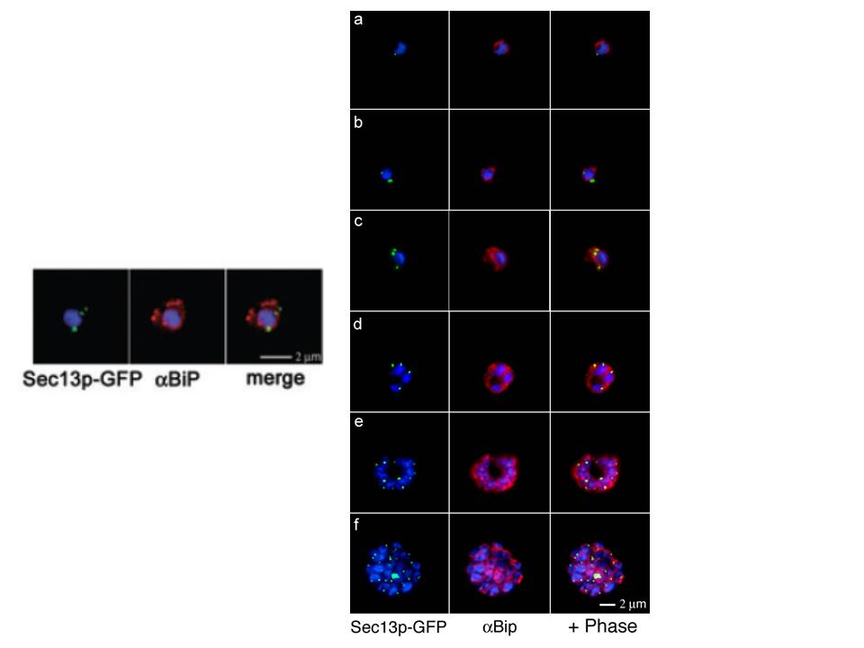
Right: Comparative analyses of transitional ER (tER) dynamics (PfSec13p) with the ER throughout the asexual life cycle of P. falciparum. Transgenic parasites expressing Sec13p–GFP (green) were fixed and stained with antibodies against the ER marker BiP (red). In ring-stage parasites (8-16 hours post invasion) GFP fluorescence is restricted to a tightly defined area in close proximity to the nucleus (a-b). A multiplication of the Sec13p–GFP-defined ER exit site takes place prior to nuclear division (24 hours post invasion, c). As the parasite matures nuclear division commences and the ER forms a more complex network, which is accompanied by further tER site multiplication (d-f).
Left: Colocalization of Sec13p–GFP (green) with the ER marker BiP (red) in fixed parasites. Antibodies against the luminal chaperone visualize the ER as a nuclear envelope with some protrusions. Sec13p–GFP is restricted to defined regions within the ER (merged image). All images show the nucleus in blue (DAPI).
Struck NS, Herrmann S, Schmuck-Barkmann I, de Souza Dias S, Haase S, Cabrera AL, Treeck M, Bruns C, Langer C, Cowman AF, Marti M, Spielmann T, Gilberger TW. Spatial dissection of the cis- and trans-Golgi compartments in the malaria parasite Plasmodium falciparum. Mol Microbiol. 2008 67:1320-30.
Other associated proteins
| PFID | Formal Annotation |
|---|---|
| PF3D7_1230700 | protein transport protein SEC13 |