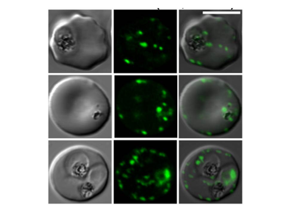
Confocal fluorescence microscopy images of transfected 3D7 P. falciparum-infected RBCs expressing a GFP chimeras directed to the Maurer’s clefts and red blood cell membrane. DIC image, the GFP fluorescence signal and an overlay of a transfectant expressing a fragment of P. falciparum erythrocyte membrane protein-1 in a chimeric construct: K119-EMP1-GFP. Scale bar = 5 µm. In trophozoite stage parasites K119-PfEMP1-GFP is associated with the Maurer’s clefts (top and middle rows). In more mature stages a weak rim signal is observed consistent with transfer of some of the K119-PfEMP1-GFP to the RBC membrane.
Tilley L, McFadden G, Cowman A, Klonis N. Illuminating Plasmodium falciparum-infected red blood cells. Trends Parasitol. 2007 23:268-77.
PubMed Article: Illuminating Plasmodium falciparum-infected red blood cells