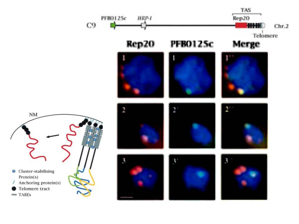
Top: Diagram showing the location of the FISH probes in chromosome 2. Bottom: Two-colour fuorescence in situ hybridization (FISH) analysis in C9 and C9-H12 asexual stages. FISH was performed as described in Materials and methods, using Rep20 as marker for chromosome end clusters (in red, panels 1-3) and as a single copy gene marker present in chromosome 2 (in green, panels 1’-3’). DNA is counterstained with 4’,6-diamidino-2-phenylindole (DAPI) (in blue). Rep20, a large telomere adjacent repetitive element, was used as a marker for the PFB0125c positioning of the telomere clusters in the nucleus. Hybridization with PFB0125c, a chromosome 2-specific marker showed one fluorescent spot located at the periphery of the nucleus.
Bottom left: Model for the spatial organization of chromosome ends in the nucleus. It predicts a distinct role for telomeres and TASs in the nuclear architecture. It may be that that perinuclear localization of chromosome ends is mediated by putative `anchoring proteins', which bind to the telomere and anchor it to nuclear membrane structural elements. TASs play an important role in the physical clustering of chromosome ends perhaps through interactions with specific cluster-stabilizing proteins, which could stabilize the association of chromosome ends. NM, nuclear membranem
Figueiredo LM, Freitas-Junior LH, Bottius E, Olivo-Marin JC, Scherf A. A central role for Plasmodium falciparum subtelomeric regions in spatial positioning and telomere length regulation. EMBO J. 2002 21:815-24.
Other associated proteins
| PFID | Formal Annotation |
|---|---|
| PF3D7_0202600 | nucleic acid binding protein, putative |
| Telomere | Telomere |