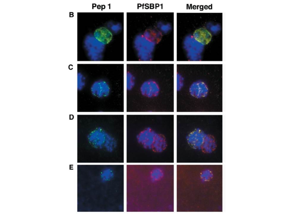
Colocalization studies with anti-peptide 1 antibodies and fixed 3D7 pRBCs (late trophozoites and schizonts). Parasite nuclei were stained with DAPI (blue). Double staining was carried out with MAb 61.2 against a 52-kDa rhoptry protein (red) and anti-peptide 1 serum (green) (A) and with mouse serum (B28) against the 77-kDa carboxy terminus of MC protein PfSBP1 (red) and anti-peptide 1 serum (STEVOR; green) (B to E). Schizonts progressd from binucleate to multinucleate in panels B to E, as determined by staining of nuclear material. Anti-peptide 1 serum was detected by using FITC conjugated goat anti-rabbit IgG. Colocalizing antibody was detected by using Texas red-conjugated goat anti-mouse IgG. Magnification, x1,000. Antibodies against STEVOR recognize proteins of the expected size (37 kDa) and localize STEVOR in Maurer’s clefts, unique membranous structures located in the cytoplasm of infected erythrocytes.
Kaviratne M, Khan SM, Jarra W, Preiser PR. Small variant STEVOR antigen is uniquely located within Maurer's clefts in Plasmodium falciparum-infected red blood cells. Eukaryot Cell. 2002 1:926-35.
Other associated proteins
| PFID | Formal Annotation |
|---|---|
| STEVOR | STEVOR |