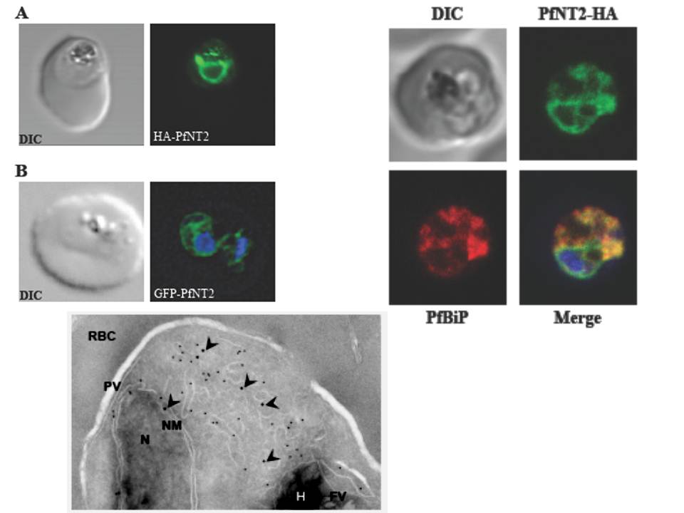
Upper left: Immunolocalization of HA-PfNT2 and GFP-PfNT2. Anti-HA (A) or anti-GFP (B) antibody (green) localizes to a compartment within transgenic parasites expressing either HA-PfNT2 (A) or GFP-PfNT2 (B). The differential contrast image (DIC) images show the location of the parasites within the erythrocyte. The nucleic acids of the GFP-PfNT2 parasites were visualized using Hoescht 33258 (blue). The perinuclear localization observed in these parasites suggested an endoplasmic reticulum location of PfNT2.
Upper right: Immunolocalization of carboxy-terminal HA-tagged PfNT2 (PfNT2-HA) to the endoplasmic reticulum. Anti-BiP (red) colocalizes with anti-HA (green). Yellow fluorescence in merged images indicate areas of red (BiP) and green (PfNT2-HA) colocalization. The nucleic acids of the parasite were visualised using Hoescht 33258 (blue).
Lower panel: Immunoelectronmicroscopy indicates PfNT2-GFP colocalizes with the ER marker PfBiP. Transmission electron micrograph of ultrathin cryosections of the intraerythrocytic trophozoite stage of P. falciparum PfNT2-GFP transgenic parasites using anti-GFP and anti-PfBiP ntibodies. The image shows immunogold labeling of anti-GFP (18-nm gold particles; indicated with arrow heads) and anti-PfBiP (12-nm gold particles) antibodies bound to intraerythrocytic P. falciparum. N, nucleus; NM, nuclear membrane; PV, parasitophorous vacuole; RBC, red blood cell cytoplasm; FV, food vacuole; H, haemozoin.
Downie MJ, El Bissati K, Bobenchik AM, Nic Lochlainn L, Amerik A, Zufferey R, Kirk K, Ben Mamoun C. PfNT2: a permease of the equilibrative nucleoside transporter family in the endoplasmic reticulum of Plasmodium falciparum. J Biol Chem. 2010 285:20827-20833.
Other associated proteins
| PFID | Formal Annotation |
|---|---|
| PF3D7_0824400 | nucleoside transporter 2, NT2 |