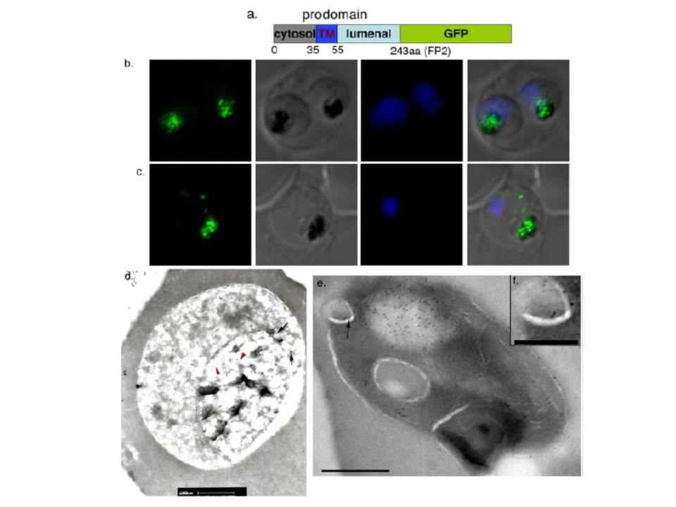
Localization of proFP-GFP wild type constructs. a, structure of the proFP2-GFP construct. Cytoplasmic (cytosol), transmembrane (TM), and lumenal portions of the prodomain are labeled. Confocal microscopic images show expression of proFP2-GFP (b) under the falcipain-2 promoter and proFP3-GFP (c) under the HSP86 promoter in young trophozoites. For this and other figures the panels show, from left to right, GFP, transmission light microscopy (black hemozoin pigment identifies the food vacuole), Hoechst staining of DNA, and a merged image. d, double immunogold labeling of an intraerythrocytic trophozoite transfected with a plasmid encoding proFP2-GFP. Native falcipain-2, labeled with antibody against the mature protease, is identified with 10-nm gold particles (red arrowheads) and GFP, labeled with anti-GFP antibody, is identified with 15-nm gold particles (black arrows). The bar indicates 0.6 mm. e, single immunogold labeling of an intraerythrocytic trophozoite transfected with proFP2-GFP and labeled with anti-GFP antibody. The arrow indicates a probable cytostome, shown in close-up in panel f.
Subramanian S, Sijwali PS, Rosenthal PJ. Falcipain cysteine proteases require bipartite motifs for trafficking to the Plasmodium falciparum food vacuole. J Biol Chem. 2007 282(34):24961-9.