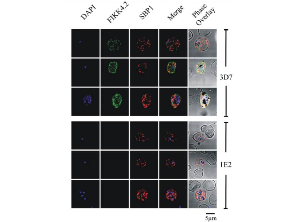
Immunofluorescence analysis of red blood cells infected with parental (3D7) or FIKK4.2 knockout (1E2) parasites. Experiments were performed on infected red blood cells that had been synchronised at either the young ring stage or the more mature trophozoite/schizont stage. Air-dried and fixed blood smears on glass slides were dual-labelled with both a-FIKK4.2 and a-SBP1 (Maurer’s cleft) antibodies. Merge and phase overlay images demonstrate clearly that FIKK4.2 and SBP1 do not co-localise in infected red blood cells. Specific punctate fluorescence was observed in the cytoplasm of RBCs infected with ring-stage 3D7 parasites. A similar pattern was observed in RBCs infected with trophozoite/schizont-stage parasites, and although the pattern of fluorescence was more diffuse, individual puncta were still clearly discernible. The puncta appeared too small and too numerous to be Maurer’s clefts, and did not co-localise with either the classical Maurer’s cleft marker SBP1.
Kats LM, Fernandez KM, Glenister FK, Herrmann S, Buckingham DW, Siddiqui G, Sharma L, Bamert R, Lucet I, Guillotte M, Mercereau-Puijalon O, Cooke BM. An exported kinase (FIKK4.2) that mediates virulence-associated changes in Plasmodium falciparum-infected red blood cells. Int J Parasitol. 2014 Feb 14. [Epub ahead of print]
Other associated proteins
| PFID | Formal Annotation |
|---|---|
| PF3D7_0424700 | serine/threonine protein kinase, FIKK family |