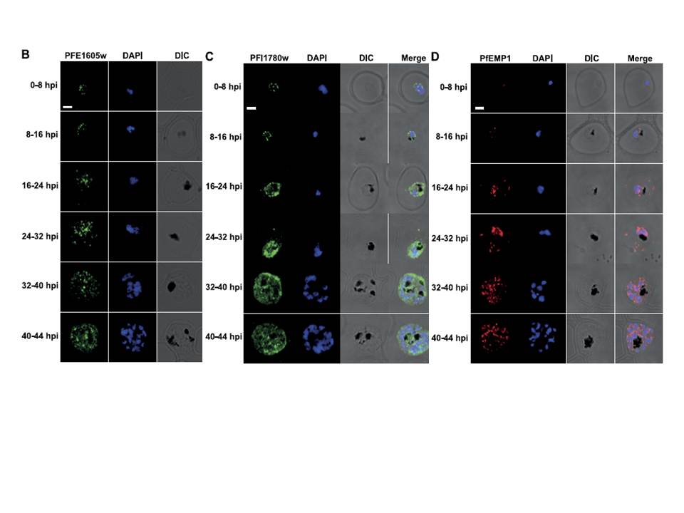
PFE1605w is cotranslocated with PfEMP1. Confocal immunofluorescence analysis of tightly synchronized 3D7 parasites. Samples were collected at 8-h intervals and stained with antibodies recognizing PFE1605w (B), PFI1780w (C), or PfEMP1 (D) and costained with DAPI. Scale bars = 2 mm. DIC, differential interference contrast microscopy. PfEMP1 is known to display a necklace of beads pattern at the parasite surface at ~8–11 hpi which was confirmed (D) and was similar to the pattern observed for PFE1605w (B). Both proteins transiently associated with Maurer’s clefts at ~16–24 hpi (D) before being transferred to the iRBC membrane, suggesting cotransport of PFE1605w with PfEMP1. In contrast to PFE1605w, PFI1780w was found within the parasite cytosol until ~24–32 hpi with limited focal fluorescence in the iRBC cytosol (C). In schizonts, PFI1780w was exported to the iRBC surface as shown in live cell imaging (B) with no distinct intermediate locations.
Oberli A, Slater LM, Cutts E, Brand F, Mundwiler-Pachlatko E, Rusch S, Masik MF, Erat MC, Beck HP, Vakonakis I. A Plasmodium falciparum PHIST protein binds the virulence factor PfEMP1 and comigrates to knobs on the host cell surface. FASEB J. 2014 [Epub ahead of print]
Other associated proteins
| PFID | Formal Annotation |
|---|---|
| PF3D7_0936800 | Plasmodium exported protein (PHISTc), unknown function |
| PfEMP1 | PfEMP1 |