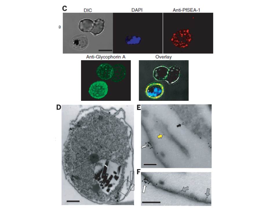
Immunolocalization of PfSEA-1. (C) Nonpermeabilized, unfixed schizont-infected RBCs were probed with mouse anti–PfSEA-1 (red) and rabbit anti–glycophorin A (green) and counterstained with DAPI to label parasite nuclei. PfSEA-1 colocalized with glycophorin A to the surface of schizont-infected RBCs. Scale bar, 5 mm. (D to F) Nonpermeabilized, unfixed schizontinfected RBCs were probed with mouse anti–PfSEA-1 (5-nm gold particles) and rabbit anti–glycophorin A (10-nm gold particles). PfSEA-1 localized to the schizont/parasitophorous vacuole membrane (black arrow), Maurer's clefts (yellow arrow), and the inner leaflet of the RBC membrane (gray arrow), whereas glycophorin A was confined to the outer leaflet of the RBC membrane (white arrow). Scale bar in (D), 0.5 mm. Scale bars in (E) and (F), 0.1 mm.
Raj DK, Nixon CP, Nixon CE, Dvorin JD, DiPetrillo CG, Pond-Tor S, Wu HW, Jolly G, Pischel L, Lu A, Michelow IC, Cheng L, Conteh S, McDonald EA, Absalon S, Holte SE, Friedman JF, Fried M, Duffy PE, Kurtis JD. Antibodies to PfSEA-1 block parasite egress from RBCs and protect against malaria infection. Science. 2014 344(6186):871-7.