Comparison of transmission electron microscopical studies on CS2 versus CS2ΔPTP1 cell line.
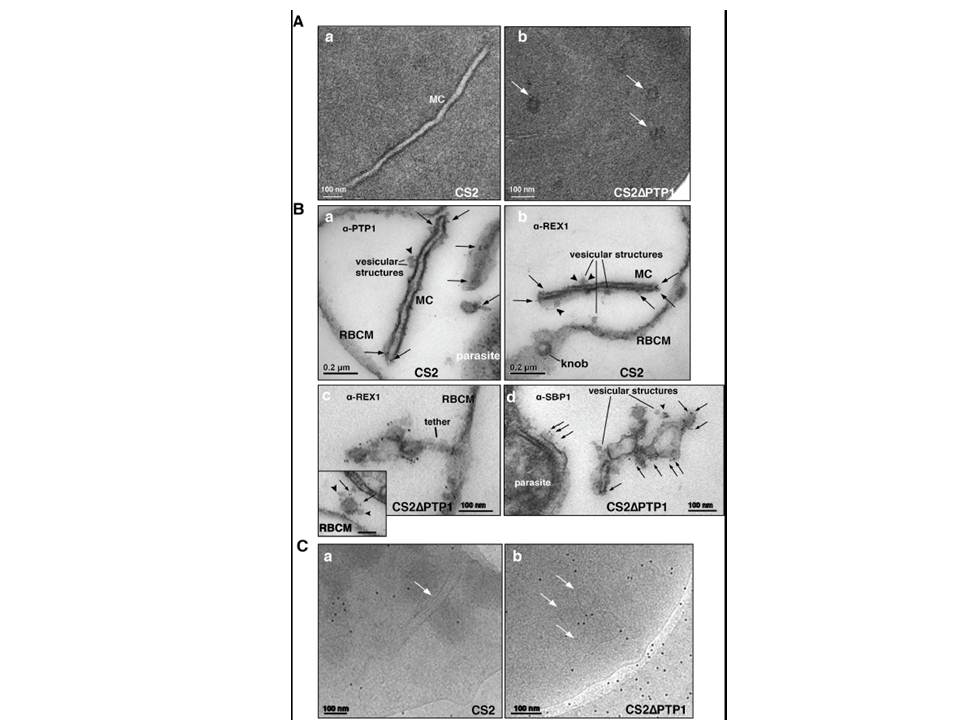
(A) Conventional chemical Fixation: a) CS2 parental line. MC (MC) display electron dense membranes and an electron lucent lumen; b) CS2ΔPTP1 cell line shows globular structures and no long lamellar MC structures (arrows). (B) Equinatoxin II treated cells with pre-embedding labelling: a) α-PfPTP1 antibodies or b) α-REX1 antibodies label the slender MC membranes (arrows) and vesicles (arrowheads) in the CS2 parasite line; c) α-REX1 or d) α-SBP1 antibodies label globular structures (arrows) and vesicular structures (arrowheads) in the CS2ΔPTP1 cell line. RBCM – red blood cell membrane. (C) The frozen hydrated samples show MC (arrow) in which the lumen looks similar to the RBC cytoplasm as described earlier. The CS2ΔPTP1 cell line shows aggregated globular structures (arrows). In CS2 typical single lamellae for MC were observed with an electron dense coat and translucent lumen (Aa). In contrast, CS2ΔPTP1 displayed electron dense areas randomly distributed through the RBC cytoplasm (Ab). The α-PfPTP1 antibodies line the MC lamella similar to REX1 localisation (B). Vesicular structures were observed that appeared to bud off MC and these had a dense lumen and less distinct membranes. These may be vesicles involved in trafficking between PVM and MC and/or MC to the RBC membrane (i.e. J-Dots). These structures were decorated with
PfPTP1 and REX1 antibodies (Ba,b). In CS2ΔPTP1 the architecture of MC was disrupted showing large aggregated globular structures (Bc,d). SBP1 and REX1 were located on these structures. Single lamellae of MC were observed in CS2 (Ca) whereas globular structures were observed for CS2ΔPTP1 (Cb).
Rug M, Cyrklaff M, Mikkonen A, Lemgruber L, Kuelzer S, Sanchez CP, Thompson J, Hanssen E, O'Neill M, Langer C, Lanzer M, Frischknecht F, Maier AG, Cowman AF. Export of virulence proteins by malaria-infected erythrocytes involves remodelling of host actin cytoskeleton. Blood. 2014 Aug 19 [Epub ahead of print]
Other associated proteins
| PFID | Formal Annotation |
|---|---|
| PF3D7_0202200 | EMP1-trafficking protein |
| PF3D7_0501300 | skeleton-binding protein 1 |