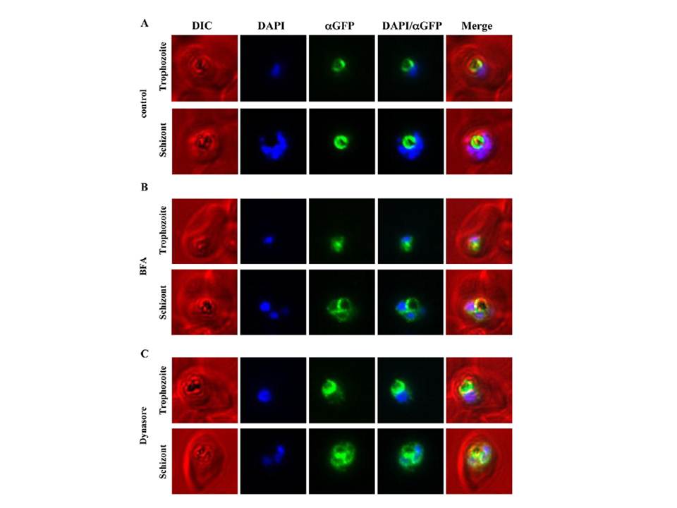
Immunofluorescence microscopy of PfCRT-GFP over-expressing parasites treated with either Brefeldin A or Dynasore. PfCRT was over-expressed as a GFP-fusion protein using an ATet-inducible expression system and treated with either BFA (5 mg/mL) for 3 h or Dynasore (40 mM/mL) for 2 h. As a control a second parasite population was treated with an equivalent volume of carrier alone (ethanol and DMSO, respectively). Following the BFA treatment, immunofluorescence microscopy was performed on fixed cells with mouse anti-GFP, Alexa-594 goat antimouse IgG and DAPI. Representative parasites in trophozoite and schizont stage are shown for control and treated parasites. (A) The control parasites show the restricted FV localisation of PfCRT-GFP (false-coloured in green). (B) BFA treated parasites show an accumulation of fluorescence around the DAPI-stained nuclei, consistent with ER localisation in addition to the FV localisation (Figure 1). (C) Treatment of parasites with Dynasore resulted in an accumulation of fluorescence around the DAPI-stained nuclei in addition to the FV membrane localisation, similar to the observed effect of BFA treatment (B).
Ehlgen F, Pham JS, de Koning-Ward T, Cowman AF, Ralph SA. Investigation of the Plasmodium falciparum food vacuole through inducible expression of the chloroquine resistance transporter (PfCRT). PLoS One. 2012;7(6):e38781.