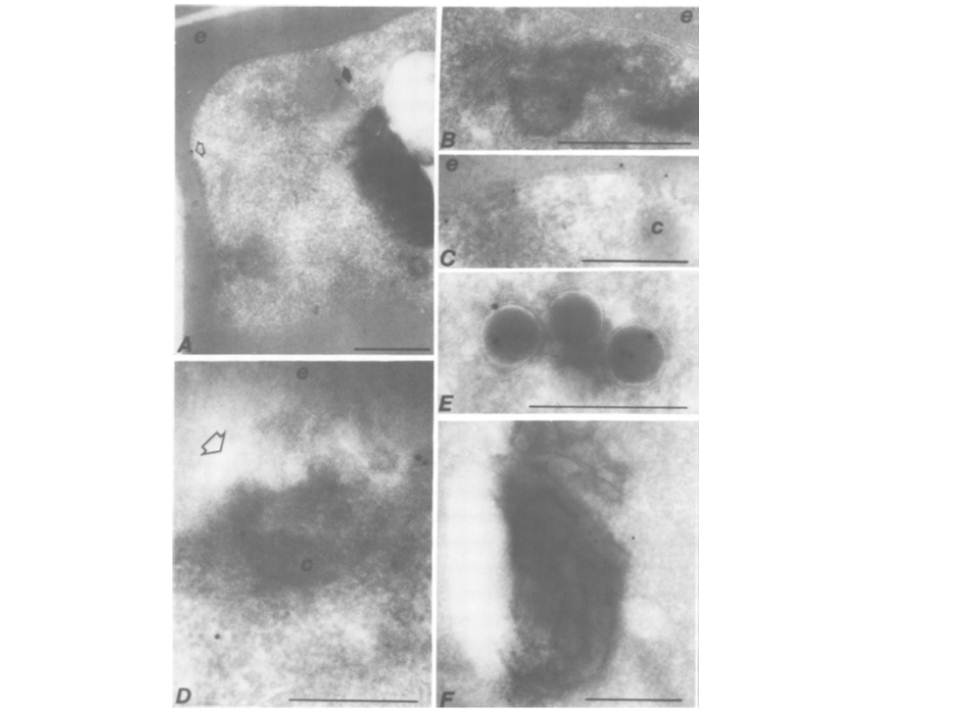
Immunoelectron microscopy of P. falciparum infected trophozoites labelled with Ab574 against falcipain I. In all micrographs 'e' signifies erythrocyte, 'c‘ signifies cytostome, and bars = 0.5mm. (A) A general view of the cell showing label associated with a hemoglobin transport vacuole (closed arrow), the surface of the parasite (open arrow), and the condensed hemozoin in the food vacuole. (B) A micrograph showing label in the vesicular omponents that subtend the surface membrane complex of the parasite. (C) A micrograph demonstrating the labeling of the pellicular membranes. (D) A section through a cytostome of the parasite (open arrow). Membrane processes are present in the lumen of the cytostome. (E) The hemoglobin transport vesicles label strongly with antibody, predominantly in the lumen of the vesicle. (F) The antibody also reacts with the digestive vacuole itself, where it is associated with the hemozoin.
Francis SE, Gluzman IY, Oksman A, Knickerbocker A, Mueller R, Bryant ML, Sherman DR, Russell DG, Goldberg DE. Molecular characterization and inhibition of a Plasmodium falciparum aspartic hemoglobinase. EMBO J. 1994 Jan 15;13(2):306-17.PubMed PMID: