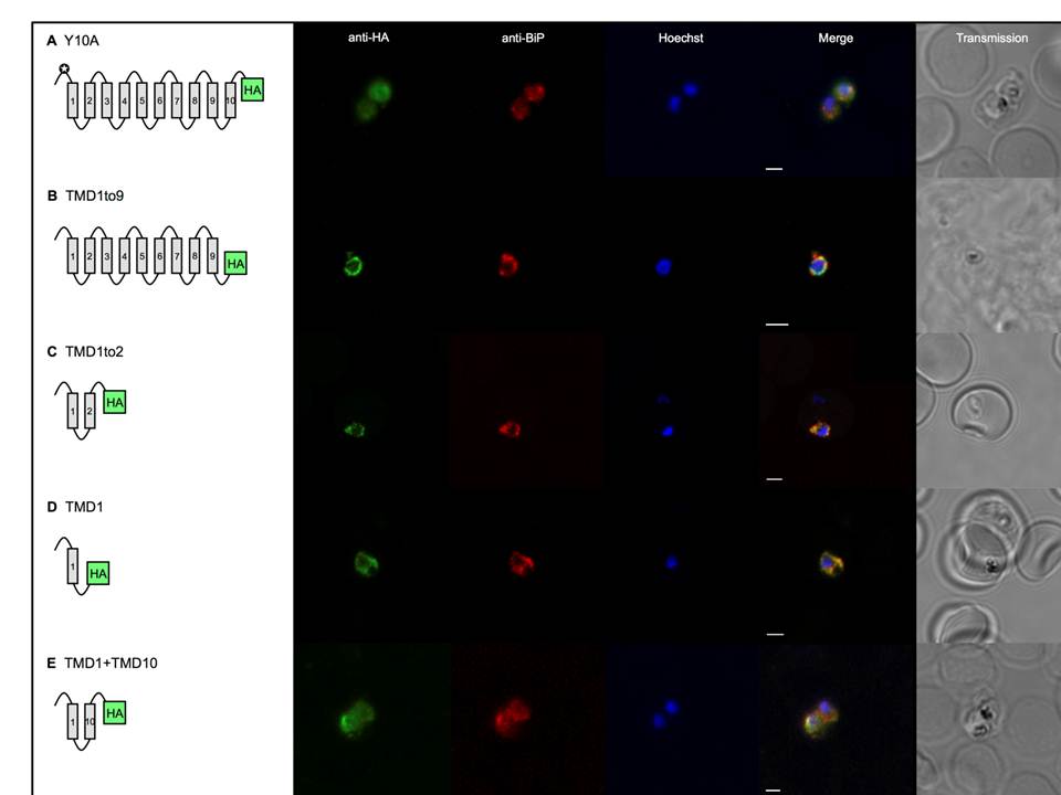
Select gene expression constructs not targeted to the apicoplast are now co-localised with the endoplasmic reticulum.
TMDs are represented by boxes and are joined by loops. All PfoTPT constructs are episomally expressed under the PfCRT promoter and tagged with triple HA at the C-terminus detected with anti-HA and secondary antibody conjugated to FITC (green). The parasite ER is detected with anti-BiP (red). Nuclei are stained with Hoechst (blue), and transmitted light images of the parasites within their host red blood cell are shown at right. Scale bars = 2μm. A. Point mutation of tyrosine residue at position 10 (✪) in the synthetic PfoTPT (Y10A) relocates protein from the apicoplast (B) to the ER, as well as the plasma. B. Removal of TMD 10 (TMD1to9), which completely abrogates targeting to the apicoplast (C), results in perinuclear localisation of the protein that almost entirely overlaps with the ER marker, BiP. C. Removal of TMDs 3, 4, 5, 6, 7, 8, 9 and 10 (TMD1to2), which completely abrogates targeting to the apicoplast (G), results in re-localisation of most of the protein to the ER. D. Removal of TMDs 2, 3, 4, 5, 6, 7, 8, 9 and 10 (TMD1), which completely abrogates targeting to the apicoplast (H), primarily results in localisation of most of the protein to the ER. E. Recombining TMD1 with TMD10 (TMD1+10) was unable to reconstitute apicoplast targeting (I) and resulted in some perinuclear ER targeting, plus some parasite plasma membrane targeting.
Lim L, Sayers CP, Goodman CD, McFadden GI. Targeting of a Transporter to the Outer Apicoplast Membrane in the Human Malaria Parasite Plasmodium falciparum. PLoS One. 2016 11(7):e0159603.
Other associated proteins
| PFID | Formal Annotation |
|---|---|
| PF3D7_1108600 | endoplasmic reticulum-resident calcium binding protein |