Saponin lysis during microscopy of a cell expressing parasitophorous vacuolar protein 2 (upper panel). The identical infected RBC is shown before (top row) and after (bottom row) lysis with saponin. release of the PV content using saponin did not abolish the signal in the parasite periphery, clearly showing that the signal around the parasite with PV2
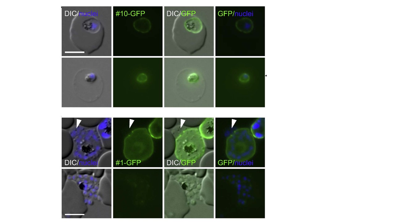
derives from the full-length protein. (B) A late schizont expressing serine/threonine protein phosphatase UIS2 (bottom panel) shows release of merozoites (arrow head) while the fluorescence still surrounds the bulk of the remainder of merozoites (top row) and no fluorescence is observed in released merozoites (bottom row). Some late stages were observed where merozoites had already been partially released but, in contrast to the merozoites in the remainder of the schizont, were not surrounded by GFP signal (arrowheads). As the released merozoites are
surrounded by PPM, this indicates that these proteins were associated
with the PVM. In agreement with this, no fluorescence was observed
with fully released merozoites.
Khosh-Naucke M, Becker J, Mesén-Ramírez P, Kiani P, Birnbaum J, Fröhlke U, Jonscher E, Schlüter H, Spielmann T. Identification of novel parasitophorous vacuole proteins in P. falciparum parasites using BioID. Int J Med Microbiol. 2017 Jul 27. [Epub ahead of print]
Other associated proteins
| PFID | Formal Annotation |
|---|---|
| PF3D7_1464600 | serine/threonine protein phosphatase uis2, putative |