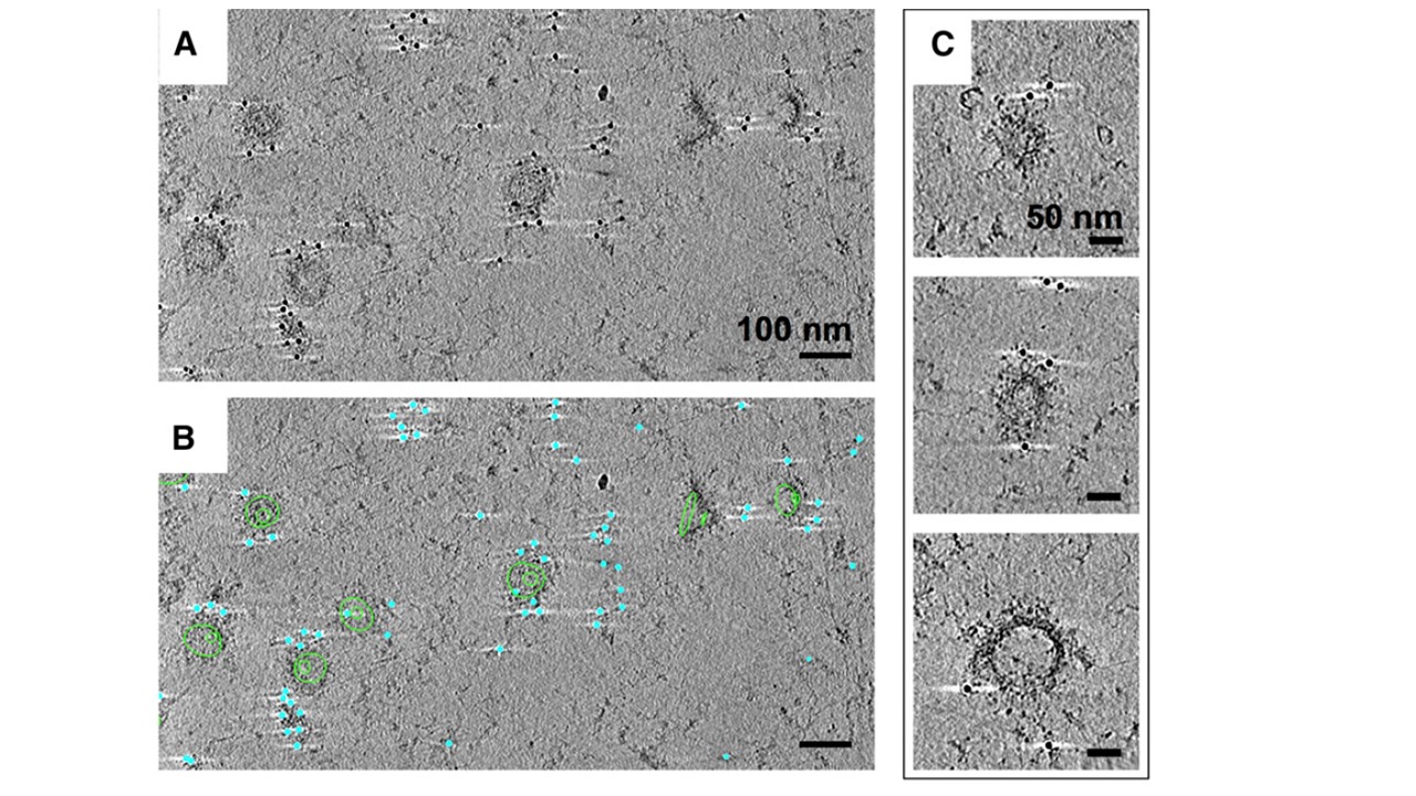
Immuno-EM labeling of KAHRP in knob skeletons. (A). Thick section (average of 50 slices) from a cryotomogram of schizont skeleton labeled with anti-KAHRP antibody 18.2 and 10-nm gold-conjugated secondary antibody. (B) The same field of view shown in (A), with 70 slices of a 3-D model overlaid, describing knob spirals by the first and last turn (green contours), and gold beads as cyan spheres. (C) Sections (30 slices each) through individual knobs in a cryotomogram of schizont skeleton labeled with mAb 18.2 and gold-conjugated secondary antibody, showing spiral structure with radiating pattern and gold labeling on the coat layer. Connected cytoskeleton is also visible. Skeletons labeled with anti-KAHRP antibody 18.2 (A-B) have gold beads clustered over the surface (C), separated from the spiral by 10 to 60 nm (Figure 6E
Watermeyer JM, Hale VL, Hackett F, Clare DK, Cutts EE, Vakonakis I, Fleck RA, Blackman MJ, Saibil HR. A spiral scaffold underlies cytoadherent knobs in Plasmodium falciparum-infected erythrocytes. Blood. 2016 127(3):343-51.