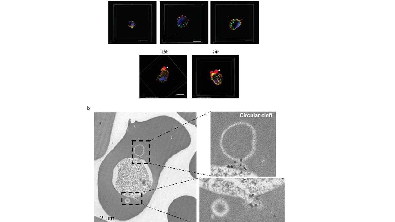
Subcellular localization of PfPV1 in ring- and trophozoite-stage parasites. (a) Results of IFA with SIM. Samples were prepared shortly after merozoite invasion as indicated. PfPV1 (green) was counter stained with EXP2 (red). Parasite nucleus was stained with DAPI (blue). Arrowheads indicate observed region of PV protrusions with accumulated EXP2. The images were made from stacks of SIM optical sections (120 nm Z-step size). Scale bars = 2 μm. (b) Results of IEM using rabbit anti-PfPV1 antibody. Gold particle labeling was observed at the base of circular clefts. Scale bar = 2 μm. EXP2 was observed in PV 1–12 h in early and mid trophozoite stages, while 18–24 h post invasion, it accumulated at a PV protrusion (a, arrowheads). Similar to other PTEX associated proteins, PfPV1 localized proximal to EXP2, and remained partially co-localized in the time course, albeit at varying degree of co-localization (a). In addition, PfPV1 also accumulated at the base of the PV protrusion in partial co-localization with EXP2 18–24 h after invasion (a; yellow signals). IEM examination of trophozoite stage parasites with the rabbit anti-PfPV1 showed accumulated gold particles at the base of circular cleft or tubular structure (b, insets).
Morita M, Nagaoka H, Ntege EH, Kanoi BN, Ito D, Nakata T, Lee JW, Tokunaga K, Iimura T, Torii M, Tsuboi T, Takashima E. PV1, a novel Plasmodium falciparum merozoite dense granule protein, interacts with exported protein in infected erythrocytes. Sci Rep. 2018 8(1):3696.
Other associated proteins
| PFID | Formal Annotation |
|---|---|
| PF3D7_1129100 | parasitophorous vacuolar protein 1 |