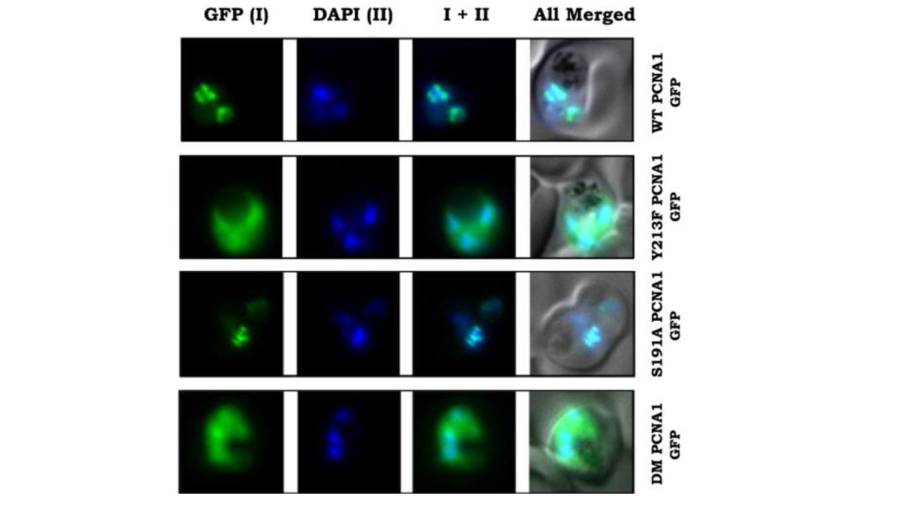
Sub-cellular localization of over-expressing GFP fusion WT and mutant PfPCNA1 in transgenic parasite lines by live cell imaging and subcellular fractionation. DAPI shows the nucleus. P. falciparum parasites over-expressing mutant PfPCNA1 as GFP fusion proteins to assess their cellular localization by live cell imaging (Supplementary Figure S2A-C, Figure 3A). These parasite lines did not show any apparent phenotypic changes compared to the untransfected 3D7 parasites. Interestingly, majority of the parasites expressing Y213F-PCNA1-GFP showed diffused GFP signals distributed all over the parasite, while parasites expressing S191A-PCNA1-GFP showed nuclear foci in many cells. Again, most parasites showed diffused GFP signals upon DM-PCNA1 expression against distinct nuclear foci of WT-PCNA1-GFP.We also calculated the number of parasites bearing PCNA1 foci.
Banu K, Mitra P, Subbarao N, Dhar SK. Role of tyrosine residue (Y213) in nuclear retention of PCNA1 in human malaria parasite Plasmodium falciparum. FEMS Microbiol Lett. 2018 Jul 19.