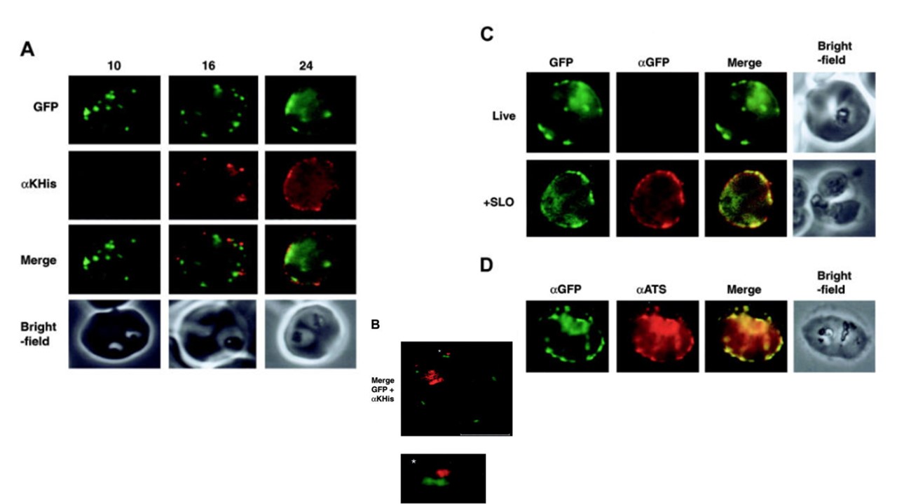
Expression of a protein domain on the surface of P falciparum–infected RBCs. All panels display parasites expressing the 3D7-K119TmATS chimeric protein. The images are from synchronized cultures of transfectants at 10, 16, and 24 hours after invasion. (A) First row, GFP fluorescence; second row, live cells incubated with αKAHRP-His (αKHis) antibodies showing appearance of the KAHRP domain on the surface 16 hours after synchronization; third row, merge of rows 1 and 2; fourth row, bright-field images. (B) Confocal images of live 3D7-K119TmATS parasites 24 hours after invasion incubated with αKAHRP-His antibodies. The image shows an overlay of the GFP fluorescence (green) and αKAHRP-His labeling (red) for a single z section (0.3 μm). The bar equals 4 μm. The asterisk indicates the adjacent locations of GFP on the inside and KAHRP-His on the outside of the RBC plasma membrane, with an enlarged image below. The internal hemozoin-associated fluorescence represents partial reflection of the excitation beam and not labeling with the antibodies. (C) Control experiments examining the specificity of antibody labeling of intact cells. First column, GFP fluorescence; second column, αGFP; third column, overlay of fluorescence images; fourth column, brightfield image. The top row shows intact parasitized RBCs with no access of the αGFP to the GFP domain inside the cell. The bottom row represents a SLO-permeabilized cell where the antibody gains access to the domain on the inside of the RBC. (D) Formaldehyde-fixed parasites show accessibility to αATS and αGFP antibodies.
Knuepfer E, Rug M, Klonis N, Tilley L, Cowman AF. Trafficking of the major
virulence factor to the surface of transfected P. falciparum-infected
erythrocytes. Blood. 2005 105(10):4078-87.