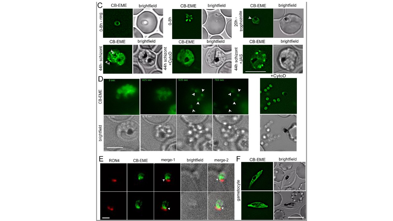
CB-EME labels actin filaments throughout the 48 h asexual life cycle with, in ring stages (0-8 h), trophozoite stages (20 h) and in 44h schizont stages. White arrows mark structures, in all likelihood F-actin, which disappear upon cytochalasin-D treatment (+CytoD), and islands of F-actin are stabilised upon jasplakinolide treatment (+JAS). Scale bar 5μm. D. Time lapse imaging of schizonts undergoing egress from the host cell, reveals a bright fluorescent signal (CB-EME) of F-actin (white arrows) at a polar end of the merozoite, appearing immediately after host cell rupture (occurs in 56%±9% egressed merozoites, N=260 from 3 independent experiments). Cytochalasin-D treatment completely prevents the polar polymerisation of F-actin in all cells (+CytoD). Scale bar 5μm. E. IFA of invading merozoites with the junction marker RON4 shows CB-EME staining close to the RON4 stain, implying that F-actin polymerises at the apical end prior to invasion. Scale bar 1 μm. F. The F-actin network and dynamics can be visualised in gametocytes. Brightfield images provided in greyscale alongside. Scale bar 5μm.
Felix Stortz J, Del Rosario M, Singer M, Wilkes JM, Meissner M, Das S. Formin-2 drives polymerisation of actin filaments enabling segregation of apicoplasts and cytokinesis in Plasmodium falciparum. Elife. 2019 8. pii: E49030.
Other associated proteins
| PFID | Formal Annotation |
|---|---|
| PF3D7_1116000 | rhoptry neck protein 4 |