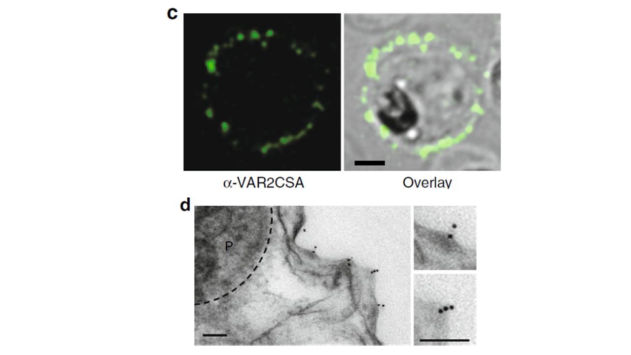
C Representative confocal immuno-florescence image of a live HbAA erythrocyte infected with G6 stained with the Zenon-labeled monoclonal antibody PAM 8.1 that is specific to the DBL3-X domain of VAR2CSA. A mid-sectional plane is shown. Bar, 2 μm. Immunofluorescence assays of live-infected erythrocytes demonstrated surface labeling, using a monoclonal antibody specific to the DBL3-X domain of VAR2CSA (c). D A representative immuno-electron micrograph of G6 showing labelling of knobs with the anti-VAR2CSA monoclonal antibody followed by a goat anti human antibody coupled to 10 nm protein A gold. Bar, 100 nm. Immuno-electron microscopy, using the same monoclonal antibody, provided further evidence of surface-presented, knob-associated VAR2CSA (d). The knobs displayed by G6 were comparable in terms of diameter and density with those found in erythrocytes infected with the parental FCR3 strain.
Sanchez CP, Karathanasis C, Sanchez R, Cyrklaff M, Jäger J, Buchholz B, Schwarz US, Heilemann M, Lanzer M. Single-molecule imaging and quantification of the immune-variant adhesin VAR2CSA on knobs of Plasmodium falciparum-infected erythrocytes. Commun Biol. 2019 May 8;2(1):172.