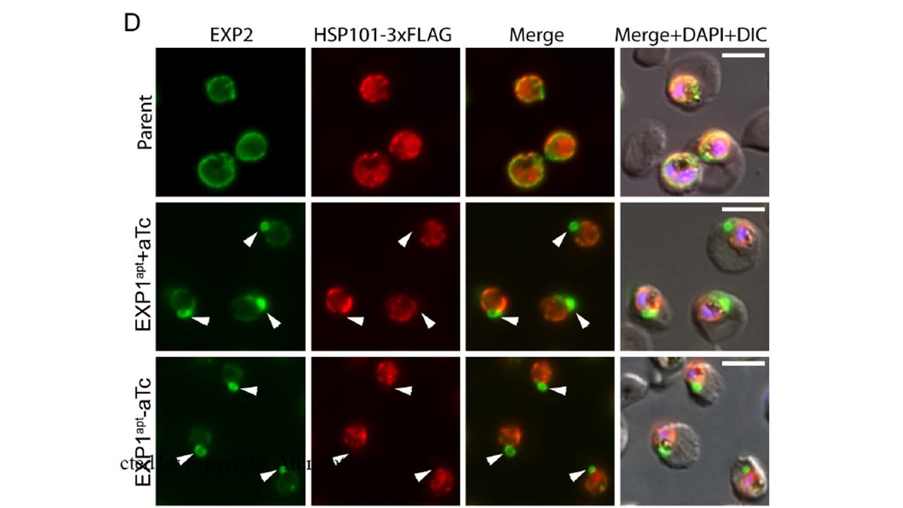
Immunofluorescence assay of parental NF54attB::HSP101-3xFLAG grown without aTc and EXP1apt grown with or without aTc for 48 hours. Arrowheads indicate areas of concentrated EXP2 signal. Scale bars are 5 μm. Data are representative of three independent experiments. EXP2 distribution was similarly altered in EXP1apt::EXP2-mNG parasites grown with or without aTc, likely reflecting the ~90% baseline reduction in EXP1 levels achieved by modification of the exp1 locus with the TDA system. A similar result was observed by fixed immunofluorescence analysis of untagged EXP2 in EXP1apt, indicating this phenomenon was not a consequence of the bulky mNG tag on EXP2. Interestingly, HSP101 did not co-localize with sites of concentrated EXP2 signal that often appeared as loops projecting away from the parasite periphery (arrowheads).
Nessel T, Beck JM, Rayatpisheh S, Jami-Alahmadi Y, Wohlschlegel JA, Goldberg DE, Beck JR. EXP1 is required for organization of EXP2 in the intraerythrocytic malaria parasite vacuole. Cell Microbiol. 2020 Jan 28. PMID: 31990132
Other associated proteins
| PFID | Formal Annotation |
|---|---|
| PF3D7_1116800 | heat shock protein 101 chaperone protein ClpB2 |