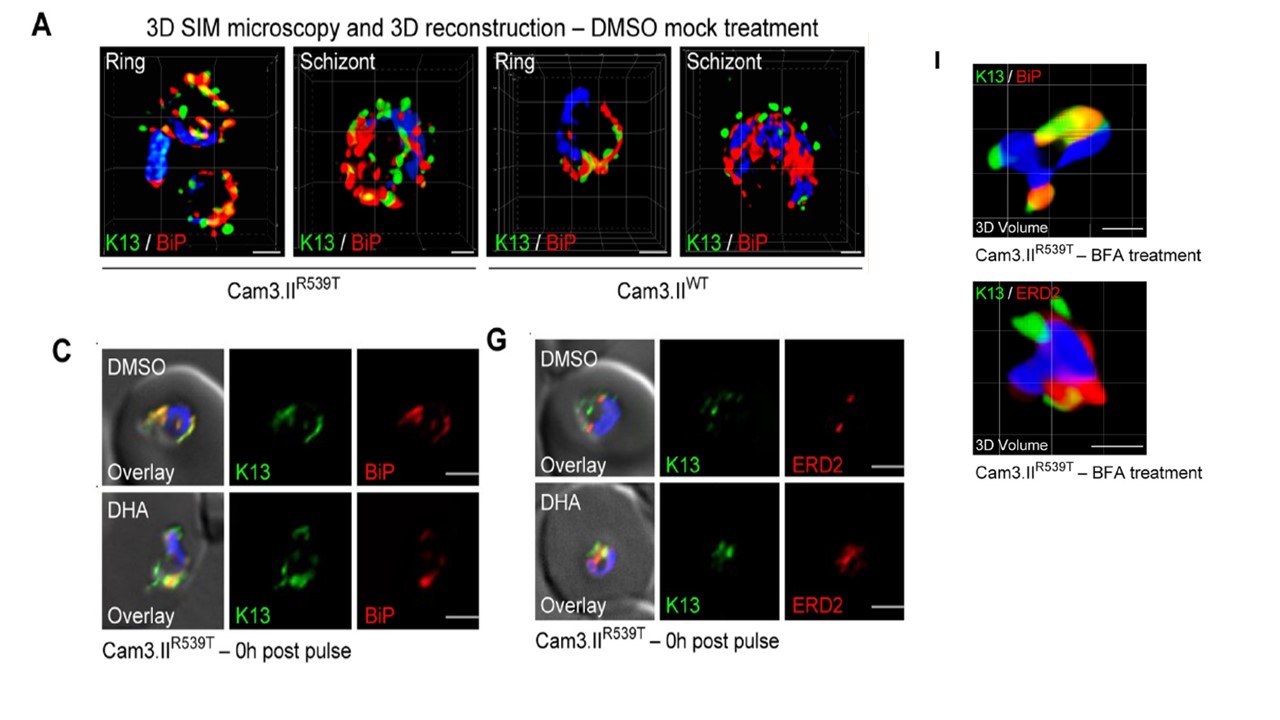
K13 shows substantial co-localization with the ER chaperone BiP but not with the cis-Golgi marker ERD2. (A) 3D SIM microscopy images showing Cam3.IIR539T or Cam3.IIWT parasites co-stained with the K13 E3 mAb (green) and antibodies to the ER chaperone BiP (red). SIM: structured illumination microscopy. Scale bars: 1 μm. (C) PCC values quantifying co-localization between K13 and BiP at 0h post DHA (6h, 700 nM) pulse, alongside representative IFA images. DMSO was used as a vehicle control. Assays were conducted using the Cam3.IIR539T and
Cam3.IIWT lines. Parasites were co-stained with the K13 E3 mAb and antibodies to BiP. (G) PCC values quantifying co-localization between K13 and ERD2 at 0h post DHA pulse, alongside representative IFA images. Parasites were co-stained with the K13 E3 mAb and antibodies to ERD2. (I) 3D volume reconstruction of deconvolved Z-stacks (15 image stacks, step size of 0.2 μm) of Cam3.IIR539T ring-stage parasites treated with BFA and co-stained with anti-K13 mAb E3 and anti-BiP (top) or anti-ERD2 (bottom). Scale bars: 1 μm.
Gnädig NF, Stokes BH, Edwards RL, Kalantarov GF, Heimsch KC, Kuderjavy M, Crane A, Lee MCS, Straimer J, Becker K, Trakht IN, Odom John AR, Mok S, Fidock DA. Insights into the intracellular localization, protein associations and artemisinin resistance properties of Plasmodium falciparum K13. PLoS Pathog. 2020 Apr 20;16(4):e1008482.
Other associated proteins
| PFID | Formal Annotation |
|---|---|
| PF3D7_1343700 | kelch protein K13 |
| PF3D7_1353600 | ER lumen protein retaining receptor |