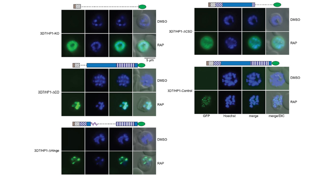
Subcellular localization of PfHP1 truncation mutants. Representative live-cell fluorescence images showing the localization of the PfHP1-GFP fusions in DMSO- and RAP-treated 3D7/HP1-KO, 3D7/HP1-DCD, 3D7/HP1-DHinge, 3D7/HP1-DCSD, and 3D7/HP1-Control lines at late schizont stage (LS [40 to 48 hpi]; generation 1, 40 h after RAP treatment). Nuclei were stained with Hoechst dye. DIC, differential interference contrast. Scale bar, 5mm. we observed that the PfHP1DCD-GFP and PfHP1DHinge-GFP fusion proteins localized to the nucleus. The PfHP1DCSD-GFP protein exhibited reduced nuclear staining, and a substantial fraction localized to the cytoplasm. Thus, the C-terminal polypeptide encompassing the CSD (amino acids 191 to 266) is required for efficient targeting of PfHP1 to the nucleus. Consistent with this observation, the small fusion protein expressed in the 3D7/HP1-KO line, which contains only the first 29 amino acids of PfHP1 fused to GFP, localized to the cytoplasm.
Bui HTN, Passecker A, Brancucci NMB, Voss TS. Investigation of Heterochromatin Protein 1 Function in the Malaria Parasite Plasmodium falciparum Using a Conditional Domain Deletion and Swapping Approach. mSphere. 2021 6(1):e01220-20..